")

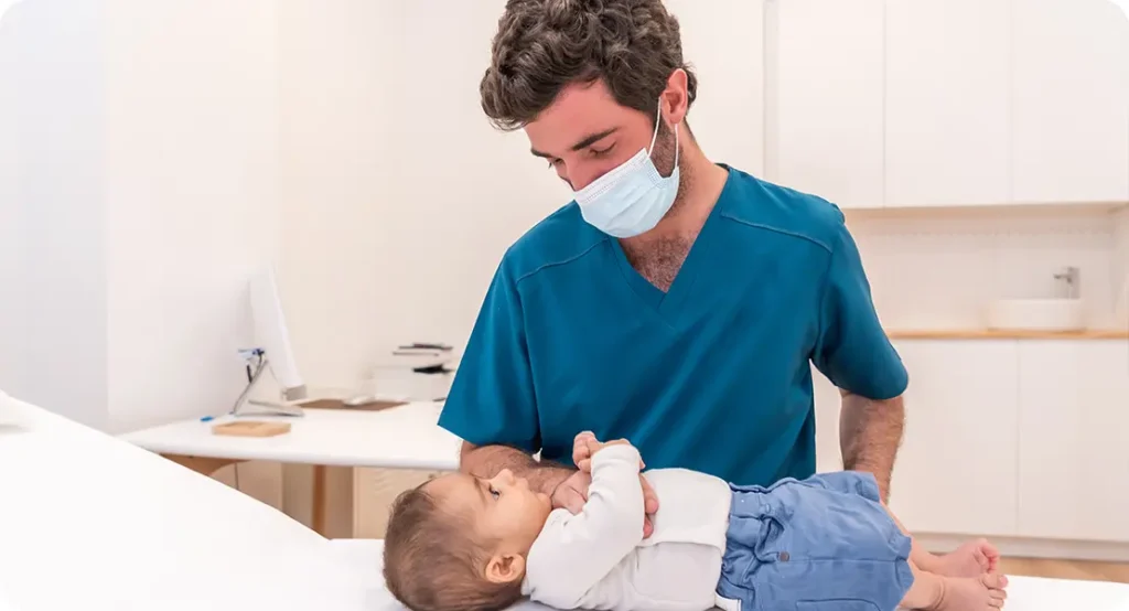
Infantile haemangiomas are the most common vascular tumours seen in infancy, typically appearing as bright red or bluish marks on a baby’s skin. These benign growths are composed of blood vessels and often emerge within the first few weeks of life. While many haemangiomas are harmless and tend to resolve naturally over time, others can grow rapidly or occur in sensitive areas, such as around the eyes, lips, or airway, potentially leading to complications. In such cases, timely medical intervention becomes essential to prevent long-term issues, both functional and cosmetic.
For many years, treatment options for haemangiomas were limited and often carried significant risks. Therapies such as systemic corticosteroids and surgical excision came with numerous side effects and uncertain outcomes, leading to a conservative approach in most cases. However, the past two decades have seen a transformative shift in how these vascular lesions are managed. Breakthroughs in medical research have introduced safer, more targeted, and less invasive treatment options that are better tolerated and more effective.
This article explores the remarkable progress in treating infantile haemangiomas, from the early use of steroids to the game-changing introduction of propranolol and the increasing role of laser therapy. We will also highlight the latest research trends, including clinical trials and genetic insights, that promise a more personalised and effective approach to managing these common childhood tumours. Whether you’re a concerned parent or a medical professional, this comprehensive guide offers valuable insights into the evolving landscape of haemangioma treatment.
1. Recognising Infantile Haemangiomas
Infantile haemangiomas are benign growths made up of blood vessels that typically develop within the first few weeks of life. These tumours can vary greatly in size, depth, and appearance, depending on their location and growth pattern. Superficial haemangiomas are often bright red, while deeper lesions may appear bluish under the skin.
Although most haemangiomas are harmless and go away over time, some may grow rapidly or form in sensitive areas, such as near the eyes, mouth, or airway. These cases require medical evaluation to prevent complications. Early identification helps in planning the most suitable treatment approach.
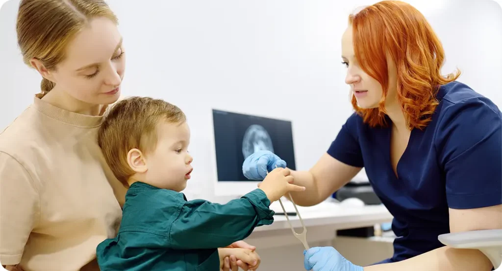
Diagnosis is generally made through clinical examination, but in complex cases, imaging techniques like ultrasound or MRI may be used. Imaging helps to assess the depth and involvement of underlying structures. Accurate diagnosis is key to ensuring safe and effective treatment.
2. Natural Progression of Haemangiomas
Haemangiomas follow a characteristic lifecycle with three phases: growth, rest, and regression. During the growth phase, typically within the first 6 to 12 months of life, the lesion expands rapidly. Parents may notice the mark increasing in size or changing in colour during this period.
After this phase, the haemangioma enters a plateau stage where growth stabilises. Although no significant changes occur, ongoing observation is necessary. This period allows clinicians to assess whether intervention is required.
The final phase is involution, where the lesion gradually shrinks and fades, often over several years. While many haemangiomas resolve without leaving a trace, some may result in residual skin changes. These changes may include scarring, skin thinning, or discoloration.
3. Early Treatment Approaches
Historically, corticosteroids were used as the primary treatment for problematic haemangiomas. These medications helped reduce inflammation and vascular growth but came with potential side effects, such as suppressed immune function and delayed development. Doses were carefully calculated to balance benefits with risk.
For more severe or life-threatening cases, chemotherapy agents like vincristine and interferon-alpha were introduced. However, these treatments were often associated with significant toxicity and only used when absolutely necessary. The risks involved made them unsuitable for mild or moderate haemangiomas.
Surgery was considered for selected cases but carried risks such as scarring, bleeding, or damage to nearby tissues. As a result, most haemangiomas were managed conservatively, even when they caused functional or cosmetic issues. The need for safer and more effective treatments became increasingly evident.
4. The Introduction of Propranolol
In 2008, propranolol emerged as a game-changer in haemangioma treatment. Originally used for heart conditions, it was found to shrink haemangiomas significantly. This discovery marked a major breakthrough in paediatric dermatology.
Propranolol works by narrowing blood vessels, reducing blood flow, and blocking signals that promote abnormal growth. Its rapid action and minimal side effects make it a preferred first-line therapy. Clinical outcomes improved dramatically after its introduction.
The shift to propranolol reduced reliance on steroids and invasive procedures. Parents and clinicians embraced the therapy due to its convenience and safety. Its effectiveness has been validated by numerous global studies.
5. Propranolol Dosage and Monitoring
Propranolol is typically started at a dose of 1 to 3 mg/kg/day, divided into two or three doses. The starting dose is often given in a hospital to monitor vital signs. This ensures the infant tolerates the medication well.
Common side effects include sleep disturbances, cold extremities, or low blood sugar. Monitoring during the early phase helps detect and manage these issues. Gradual dose adjustments are made based on weight and response.
Follow-up appointments are scheduled every few weeks to track progress. Most haemangiomas respond within days to weeks of starting treatment. The therapy usually continues for 6 to 12 months.
6. Use of Topical Beta-Blockers
Topical timolol is used for small, superficial haemangiomas. It’s especially helpful for lesions on the face where oral medication may not be necessary. The solution or gel is applied directly to the skin.
Timolol works similarly to propranolol but with lower systemic absorption. This reduces the risk of serious side effects. It’s often the first choice for uncomplicated cases.
Monitoring is still important, especially in infants under six months. Even topical medications can occasionally cause systemic reactions. A doctor’s guidance ensures safe usage.
7. Laser Therapy for Haemangiomas
Laser therapy is used to treat haemangiomas that cause ulcers or fail to regress fully. The most common type used is the pulsed dye laser (PDL). It targets blood vessels without damaging surrounding tissue.
Laser treatment can reduce redness, flatten raised areas, and accelerate healing. It’s often used alongside propranolol for enhanced results. Sessions are spaced several weeks apart.
Side effects include temporary swelling, bruising, or pigment changes. Most effects are mild and resolve on their own. Laser is a safe, effective adjunct for select cases.
8. Choosing the Right Laser Type
The pulsed dye laser (PDL) is preferred for superficial lesions. It offers precise targeting of blood vessels with minimal discomfort. It’s the standard choice for early and flat haemangiomas.
For deeper or resistant lesions, Nd:YAG or diode lasers may be used. These penetrate further but also carry higher risks. Expert assessment is needed to select the right modality.
Laser parameters are adjusted based on skin type, lesion depth, and patient age. This personalised approach ensures safety and results. Sessions are usually performed in outpatient settings.
9. Combining Therapies for Best Results
Many haemangiomas benefit from a combination of treatments. Oral propranolol can reduce lesion size, while laser addresses residual discolouration. Topical timolol adds further support for superficial areas.
Using multiple therapies allows for tailored treatment plans. Combination therapy improves both speed and quality of results. It’s especially helpful in mixed or stubborn lesions.
Coordination between dermatologists and paediatricians ensures optimal outcomes. A multidisciplinary approach enhances patient safety and family satisfaction. Timely intervention prevents complications.
10. Side Effects and Safety Considerations
While modern therapies are generally safe, side effects can still occur. With propranolol, the main risks include low heart rate, low blood sugar, or wheezing. These are rare but require careful monitoring.
Laser therapy may cause redness, crusting, or temporary pigment changes. Most of these are mild and resolve quickly. Proper aftercare helps minimise discomfort.
Parents should be educated on signs of adverse reactions. Clear communication improves adherence to treatment plans. Overall, the safety profile of current therapies is excellent.
11. Monitoring Progress and Adjusting Treatment
Regular monitoring is crucial during haemangioma treatment. Follow-up visits help track changes in size, colour, and texture. These assessments guide further decisions about therapy.
Parents are often advised to take regular photographs for comparison. This visual documentation can be very useful in clinical evaluations. Adjustments in dose or therapy type may be made based on progress.
If the haemangioma responds poorly or new symptoms arise, a different treatment approach may be needed. Timely updates help prevent complications. Continuous follow-up is a key element of successful care.
12. Addressing Ulcerated Haemangiomas
Some haemangiomas develop painful ulcers, particularly in high-friction areas. These require specialised wound care to prevent infection. Ulceration may also be a reason to start or intensify treatment.
Topical antibiotics, dressings, and barrier creams are commonly used. Pain management may be necessary depending on the severity. Healing often improves once systemic treatment is started.
Laser therapy can also help in some ulcerated cases. By sealing the vessels, it promotes quicker healing. Prompt intervention ensures better recovery.
13. Psychological and Social Impact
Visible haemangiomas, especially on the face, can lead to self-consciousness as children grow older. This may affect social interactions or emotional well-being. Early treatment often prevents these issues.
Parents may also feel distress, guilt, or anxiety about their child’s appearance. Open discussions with healthcare providers can be reassuring. Emotional support is an important part of holistic care.
Support groups and community forums can help families feel less isolated. Sharing experiences with others can reduce stigma. Mental health support should not be overlooked in comprehensive management.
14. When to Refer to a Specialist
While many haemangiomas can be managed by general practitioners, complex cases should be referred early. Lesions near vital structures or those growing rapidly need expert care. Timely referral improves outcomes.
Paediatric dermatologists have specialised knowledge and tools to assess and treat difficult haemangiomas. They may also have access to multidisciplinary teams. Referrals ensure safe and effective treatment planning.
Parents should never hesitate to ask for a second opinion. Early specialist involvement often prevents long-term problems. Good communication between healthcare providers benefits the patient.
15. Imaging Techniques for Deeper Lesions
When haemangiomas are not fully visible on the surface, imaging is helpful. Ultrasound is often the first step in evaluating lesion depth. It is safe, quick, and does not require sedation.
MRI may be used in more complex or extensive cases. This gives a clear view of surrounding tissues and any structural involvement. Imaging supports better treatment planning.
Sometimes imaging is repeated during treatment. This helps assess how well the lesion is responding. Accurate imaging improves decision-making.
16. PHACE Syndrome and Associated Risks
Some haemangiomas are part of a wider medical condition known as PHACE syndrome. This involves large facial haemangiomas and possible brain, heart, or eye abnormalities. Early diagnosis is essential.
PHACE syndrome is rare but requires a full evaluation by specialists. Neurologists, cardiologists, and ophthalmologists may be involved. Multidisciplinary care is key to managing all aspects.
MRI and echocardiogram are part of the diagnostic work-up. If PHACE is suspected, close follow-up is needed. Awareness of this condition helps avoid missed diagnoses.
17. Role of Genetic and Molecular Research
Recent studies suggest that genetics may influence haemangioma development. Specific gene mutations and molecular pathways are being investigated. Understanding these factors may lead to better-targeted therapies.
Research into angiogenic signalling and vascular biology continues. This helps explain why some haemangiomas behave differently. New discoveries are shaping personalised treatment plans.
As science advances, therapies may be based on individual genetic profiles. This approach could increase success rates. Genetics may hold the key to future breakthroughs.
18. Clinical Trials and Emerging Treatments
Ongoing trials are testing new haemangioma treatments. These include alternate beta-blockers, topical drugs, and biologics. Results from these studies will influence future guidelines.
Participation in trials gives patients access to promising therapies. It also contributes to medical knowledge. Specialist centres often provide information about available studies.
Early findings are promising, especially for resistant haemangiomas. Monitoring trial data helps clinicians stay informed. The field continues to evolve rapidly.
19. Accessibility and Cost of Care
Access to haemangioma treatment can vary based on geography and healthcare system. While propranolol is affordable, laser therapy may not be. Insurance coverage differs widely.
Clinics may offer financial aid or refer families to support programmes. Transparency in costs allows better planning. Outreach can help underserved communities receive care.
Ensuring equity in access is a healthcare priority. All children deserve the best available treatment. Policy support is important in bridging gaps.
20. Future Directions in Haemangioma Management
The future of haemangioma treatment lies in customisation. Therapies will increasingly be tailored to lesion type, location, and patient characteristics. Technology will support these decisions.
Telemedicine may improve follow-up and reach families in remote areas. AI tools could assist in diagnosis and monitoring. Innovation continues to drive progress.
Long-term studies will guide best practices. Collaboration across centres ensures ongoing learning. The outlook for affected children is more hopeful than ever.
Final Thoughts: A Brighter Future for Infantile Haemangioma Care
The treatment of infantile haemangiomas has advanced significantly, offering parents safer and more effective options than ever before. With the introduction of propranolol, laser therapies, and topical agents, management has become more targeted, less invasive, and highly successful in preventing complications.
As research continues and personalised care evolves, outcomes will only improve. Early recognition and timely intervention remain crucial, along with access to specialist support. Parents can take comfort in knowing that modern treatments are not only effective but also considerate of each child’s unique needs.
You can get in touch with us at The London Dermatology Centre to book a consultation with one of our expert paediatric dermatologists and explore a treatment plan designed specifically for your child’s wellbeing.
References:
- Hoeger, P.H., Harper, J.I., Baselga, E., Bonnet, D., Boon, L.M. and Frieden, I.J., 2015. Treatment of infantile haemangiomas: recommendations of a European expert group. European Journal of Pediatrics, 174(7), pp.855–865.
- Léauté-Labrèze, C., Hoeger, P., Mazereeuw-Hautier, J., Guibaud, L., Baselga, E., Posiunas, G. et al., 2015. A randomized, controlled trial of oral propranolol in infantile hemangioma. New England Journal of Medicine, 372(8), pp.735–746. Available at: https://www.nejm.org/doi/full/10.1056/NEJMoa1404710
- Drolet, B.A., Frommelt, P.C., Chamlin, S.L., Haggstrom, A.N., Bauman, N.M., Chiu, Y.E. et al., 2013. Initiation and use of propranolol for infantile hemangioma: report of a consensus conference. Pediatrics, 131(1), pp.128–140. Available at: https://pediatrics.aappublications.org/content/131/1/128
- McCuaig, C.C. and Powell, J., 2019. Laser treatment of vascular lesions in children. Dermatologic Clinics, 37(3), pp.369–380.
- Kim, K.H. and Geronemus, R.G., 2009. Pulsed dye laser treatment of infantile hemangiomas: an update. Dermatologic Surgery, 35(12), pp.2028–2034. Available at: https://doi.org/10.1111/j.1524-4725.2009.01334.x