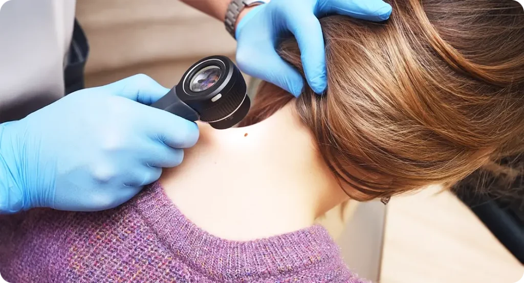
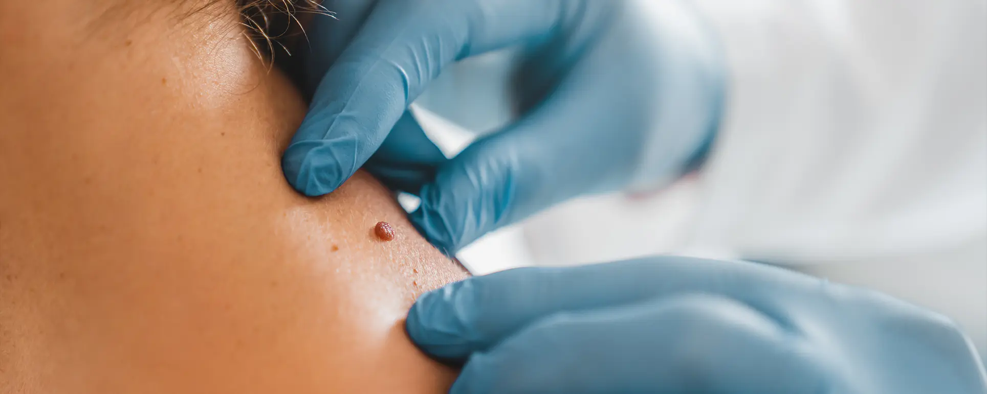
Artificial intelligence (AI) is fast becoming an integral part of modern dermatology, particularly in the monitoring and early detection of melanoma. One of the most promising applications is AI-powered mole mapping, which involves using machine learning algorithms to analyse skin images over time. This technology is designed to identify suspicious changes in moles and skin lesions with greater precision and efficiency than manual observation alone.
With skin cancer rates on the rise globally, early detection remains the cornerstone of effective treatment. AI systems aim to bridge gaps in healthcare access, improve diagnostic accuracy, and reduce human error. But how effective are these tools in real-world clinical settings? What do current studies tell us about their reliability, accuracy, and potential biases?
This article delves into the latest research on AI-assisted mole mapping to provide a balanced, evidence-based overview of its strengths, challenges, and future role in dermatology.
How AI Mole Mapping Works: Technology Behind the Tool
At the heart of AI mole mapping are advanced algorithms trained on thousands—if not millions—of dermatoscopic and clinical images. These algorithms employ deep learning models, particularly convolutional neural networks (CNNs), which are adept at recognising patterns in visual data.
By analysing features such as asymmetry, border irregularity, colour variation, diameter, and evolution (ABCDE), AI systems can flag potentially malignant lesions for further assessment. Some platforms even offer full-body mole mapping using 3D imaging, which enables comprehensive tracking of changes across the skin surface. The ability to compare historical images over time allows AI to spot subtle developments that might be missed by the human eye.
This continuous surveillance is especially useful for high-risk individuals with numerous atypical moles. Importantly, AI tools are not meant to replace dermatologists but to support them by acting as a second pair of eyes—providing rapid, scalable analysis that enhances clinical decision-making.
Accuracy and Diagnostic Value: What Studies Reveal
A growing body of research has examined the diagnostic accuracy of AI-powered mole mapping systems. A 2020 study published in Nature found that a deep learning model outperformed 58 dermatologists in distinguishing malignant melanomas from benign lesions, achieving an area under the curve (AUC) of 0.94.
Other large-scale meta-analyses have supported these findings, noting that AI tools can match or exceed the diagnostic performance of trained specialists, particularly when high-quality imaging is available. However, the real-world effectiveness of these systems depends heavily on factors such as image resolution, lighting conditions, and patient skin type. In clinical practice, false positives remain a concern, potentially leading to unnecessary biopsies and anxiety.
Conversely, false negatives—although less common—could delay critical interventions. Overall, current evidence suggests that AI tools are highly sensitive and reasonably specific, but must be used as part of a broader clinical workflow rather than in isolation.

Bias and Data Limitations: The Diversity Challenge
One of the most significant concerns surrounding AI in dermatology is algorithmic bias, particularly related to skin tone. Many AI systems have been trained predominantly on images of lighter-skinned individuals, which raises concerns about their effectiveness in diagnosing melanoma in patients with darker skin tones.
A 2021 review in JAMA Dermatology highlighted this issue, noting that underrepresentation of diverse skin types in training datasets can lead to skewed diagnostic outcomes. This disparity not only affects the accuracy of detection but also reinforces existing health inequities. Some tech developers are actively working to diversify image databases, but progress is uneven. Until these tools are validated across a broad spectrum of ethnicities, clinicians must remain cautious.
Transparency about training data and performance metrics is essential, as is regulatory oversight to ensure fairness. Bias in AI is not merely a technical flaw—it has real-world implications for patient care and trust in digital health tools.
Integration into Clinical Practice: Benefits and Barriers
AI-powered mole mapping tools are increasingly being integrated into dermatology clinics, telemedicine platforms, and even consumer apps. Their appeal lies in the potential to streamline skin checks, improve early detection, and optimise clinical resources. For dermatologists, these systems offer decision support that enhances accuracy without significantly increasing consultation time.
For patients, especially those in remote or underserved areas, AI offers improved access to preliminary assessments. However, adoption is not without challenges. High costs, data privacy concerns, lack of regulatory standards, and the need for specialised training are key barriers. Moreover, integration into existing electronic health records (EHRs) and workflow systems requires careful planning. Successful implementation depends on a hybrid model, where AI augments rather than replaces human expertise.
Encouragingly, early adopter clinics report positive experiences, citing improved efficiency and patient satisfaction. As tools become more refined, their role is likely to grow—but only with adequate safeguards and clinician buy-in.
Ethical Considerations and Patient Consent
As AI systems become more embedded in medical diagnostics, ethical issues surrounding consent, transparency, and accountability come to the fore. Patients must be clearly informed when AI is used in their diagnostic process and what it entails. Informed consent should include an explanation of the tool’s capabilities, limitations, and the role of the human clinician in interpreting results.
Another pressing concern is data security. Skin images are highly personal, and their storage, transmission, and analysis must comply with stringent data protection laws such as GDPR. Furthermore, in the event of a diagnostic error, questions of liability become complex—who is responsible, the software developer or the healthcare provider?
Addressing these questions requires interdisciplinary collaboration between ethicists, clinicians, engineers, and legal experts. As mole mapping technology evolves, so too must the frameworks that govern its use. Responsible innovation is the key to building trust and ensuring ethical adoption.
Teledermatology and Remote Monitoring: Extending Reach with AI
One of the most transformative impacts of AI mole mapping is its integration into teledermatology, offering remote skin surveillance for patients who cannot easily access in-person care. This is particularly relevant in rural areas or regions with a shortage of dermatologists. AI tools embedded in telehealth platforms allow patients to upload high-resolution images of their skin for automated analysis.
Dermatologists can then review flagged lesions and prioritise urgent cases. A 2022 study in The British Journal of Dermatology found that remote mole assessments supported by AI significantly reduced time to diagnosis for high-risk lesions. Additionally, AI can help manage ongoing surveillance for patients with numerous atypical moles by identifying subtle changes between visits.
While challenges remain in standardising image quality and ensuring secure transmission, the marriage of AI and telemedicine is expanding the frontiers of dermatological care, offering scalable, efficient, and equitable solutions for early melanoma detection.
Consumer-Facing Mole Mapping Apps: Empowerment or Risk?
As AI becomes more consumer-accessible, a growing number of mobile apps now offer mole mapping and skin analysis directly to users. These apps claim to provide immediate feedback on whether a mole appears suspicious, empowering users to take charge of their skin health. However, the clinical reliability of such tools varies significantly.
Few are regulated as medical devices, and most lack peer-reviewed validation. A 2021 review in The Lancet Digital Health raised concerns about over-reliance on these consumer tools, noting a high rate of false positives and a lack of transparency around algorithm training.
While these apps can raise awareness and prompt earlier clinical visits, they may also cause undue alarm or a false sense of security. Experts recommend that consumer apps be used as educational aids rather than diagnostic tools, and that any concerning findings be reviewed by a qualified healthcare provider.
AI in Multimodal Diagnostics: Merging Data for Better Outcomes
The future of AI mole mapping may lie in combining skin imaging with other forms of patient data to enhance diagnostic accuracy. Researchers are exploring how to integrate genetic profiles, patient history, and risk factors (such as UV exposure and family history of melanoma) into AI models. This multimodal approach allows algorithms to assess not just what a mole looks like, but also the context in which it appears.
By understanding the patient’s overall risk profile, AI systems can prioritise high-risk lesions more effectively. Early trials suggest that combining dermatoscopic images with metadata improves both sensitivity and specificity.
Such advances move AI closer to personalised dermatology—where each diagnosis is tailored not just to a lesion’s appearance, but to the patient as a whole. As these systems evolve, they may become crucial tools not just for detection but also for risk stratification and management planning.

Training the Next Generation: AI in Medical Education
AI-powered mole mapping tools are also making their way into medical education, helping to train future dermatologists. With access to vast image datasets and real-time analysis tools, medical students and junior clinicians can hone their diagnostic skills in a structured, feedback-rich environment.
Some platforms offer simulated diagnostic scenarios, allowing trainees to practise identifying lesions and receive immediate AI-generated assessments alongside expert commentary. Studies have shown that this dual approach improves learning outcomes and boosts diagnostic confidence. Furthermore, AI can help standardise education across institutions, ensuring trainees are exposed to a broad spectrum of lesion types and skin tones.
By incorporating AI into curricula, educators are not only improving diagnostic competence but also preparing clinicians to work alongside these tools in future practice. This symbiosis of human and machine learning could raise overall standards of care and diagnostic consistency across the field.
Regulatory Landscape and Future Outlook
As AI tools become more embedded in clinical practice, regulatory oversight is crucial to ensure safety, efficacy, and accountability. In the UK, the Medicines and Healthcare products Regulatory Agency (MHRA) is beginning to address how AI-driven diagnostic devices are evaluated. Clear guidance is needed on clinical validation, data transparency, and post-market surveillance.
Currently, only a few AI mole mapping platforms are approved for clinical use under rigorous regulatory frameworks. For AI tools to gain widespread trust, they must be subjected to the same standards of scrutiny as traditional diagnostic methods.
Looking ahead, advancements in AI explainability, real-time calibration, and bias mitigation will likely shape the next wave of innovations. With responsible governance and continued research, AI mole mapping has the potential to become a cornerstone of skin cancer prevention—offering early, accurate, and equitable detection for all.
Longitudinal Tracking and Change Detection
One of the most compelling advantages of AI in mole mapping is its capacity for longitudinal tracking—monitoring individual lesions over time to detect changes that may indicate malignancy. Unlike human observers, AI systems do not suffer from fatigue or recall bias, making them especially effective for serial comparisons.
Many platforms now store baseline images and employ algorithms that highlight even minute alterations in size, shape, or colour. This functionality is particularly important for patients with multiple dysplastic nevi or a personal history of melanoma, who require vigilant surveillance. A 2023 study published in Dermatology and Therapy demonstrated that AI-enhanced monitoring detected changes in high-risk lesions an average of 3.5 months earlier than standard visual checks alone.
Such early warnings can be life-saving, leading to prompt excision and treatment. By supporting clinicians with detailed visual timelines, AI mole mapping tools are not just diagnostic aids—they are also powerful tools for preventative care.
Cost-Effectiveness and Resource Allocation
While the initial investment in AI mole mapping systems can be high, several health economic analyses suggest that these tools may be cost-effective in the long term. By streamlining diagnostics and reducing unnecessary biopsies, AI tools can help optimise resource use across dermatology services.
A 2022 cost-utility analysis conducted in Germany found that AI-supported mole assessments resulted in lower healthcare expenditures when considering reduced dermatologist workload and earlier melanoma treatment, which often involves less aggressive and costly interventions.
Additionally, AI can help triage patients more effectively, ensuring that those at highest risk receive priority appointments. In overburdened public health systems, this can lead to faster diagnoses and reduced waiting times, improving outcomes while also controlling costs. As governments and health providers seek scalable innovations to deal with rising skin cancer rates, AI offers a promising return on investment—both in financial and human terms.
Limitations in Rare or Atypical Lesions
Despite its many strengths, AI is not infallible—particularly when it comes to rare or atypical lesions. Algorithms trained on large datasets often excel at recognising common presentations but may struggle with unusual subtypes such as amelanotic melanomas, which lack pigmentation, or nodular melanomas that do not follow typical ABCDE criteria.
Furthermore, lesions that mimic malignancy—such as seborrhoeic keratoses or angiomas—can confound even sophisticated models. A 2021 analysis in Clinical and Experimental Dermatology reported lower specificity in detecting non-classic presentations, leading to a higher rate of false positives.
This limitation underscores the necessity of clinician oversight. AI should be seen as a supportive tool rather than a definitive diagnostic engine. By recognising its boundaries, dermatologists can better leverage its strengths while compensating for its weaknesses—ensuring that no critical diagnosis is missed due to algorithmic blind spots.
Impact on Patient Anxiety and Behaviour
The psychological impact of AI mole mapping on patients is a growing area of interest. On one hand, having immediate access to AI-generated assessments can empower patients, encourage regular skin monitoring, and foster proactive health behaviours. On the other, false positives or alarming notifications from consumer-facing apps can heighten anxiety and lead to unnecessary clinic visits.
Managing expectations is key. Clinicians must help patients interpret AI results within the broader context of their individual risk profile and provide clear guidance on next steps. Some clinics have started incorporating counselling protocols alongside AI assessments to address patient concerns and ensure informed decision-making.
Research into patient perceptions of AI in dermatology remains limited, but early findings suggest that transparency, reassurance, and clinician involvement are critical for maintaining trust. As AI becomes more commonplace, understanding its psychological effects will be essential to delivering holistic, patient-centred care.
Global Access and Equity
AI-powered mole mapping has the potential to improve dermatological care not just in developed countries, but also in low-resource settings where dermatologists are scarce. With smartphone access becoming increasingly widespread, AI could help bridge healthcare gaps by offering preliminary screenings in remote regions. Non-profit organisations and global health initiatives are beginning to explore how simplified AI tools can support community health workers in early melanoma detection.
However, real-world deployment faces significant hurdles. These include poor internet connectivity, lack of digital literacy, and limited access to high-resolution cameras. Moreover, as discussed earlier, many AI tools have not yet been validated on diverse skin types, limiting their effectiveness in parts of Africa, Asia, and Latin America.
To fulfil their global promise, developers must prioritise inclusive training datasets and collaborate with local health systems to adapt tools to context-specific needs. Equity must be central to innovation if AI is to serve as a universal force for good.
Collaboration Between Dermatologists and Data Scientists

The successful development and deployment of AI mole mapping tools hinges on collaboration between clinicians and technologists. Dermatologists bring essential domain knowledge about skin pathology, while data scientists contribute expertise in algorithm design and optimisation. When these disciplines work in tandem, the result is more robust, clinically relevant tools.
Multidisciplinary teams are now common in AI health research, with many hospitals and universities forming dedicated digital dermatology units. These teams not only develop new models but also test them in clinical environments, creating feedback loops that refine performance.
Furthermore, clinician involvement in the design process helps ensure that AI interfaces are user-friendly and aligned with real-world diagnostic workflows. As the field evolves, collaborative innovation will be key to producing AI systems that are not just technically impressive but also deeply useful to the people who rely on them—patients and clinicians alike.
Conclusion: A Promising but Cautious Path Forward
Certainly. Here’s the revised version with your requested statement added:
AI-powered mole mapping holds significant promise for transforming skin cancer surveillance and early diagnosis. Studies to date demonstrate encouraging levels of accuracy, with many tools rivalling expert dermatologists in controlled settings.
However, challenges related to bias, real-world reliability, clinical integration, and ethical use must be addressed before widespread adoption. The technology is best seen as a powerful complement to clinical judgement—not a replacement.
As image databases grow more diverse and systems become more sophisticated, AI is likely to play an increasingly central role in dermatological care. For now, ongoing research, rigorous validation, and thoughtful implementation will determine whether this technology becomes a true game-changer in the fight against melanoma.
If you are interested in mole mapping and would like to discuss this with one of our expert dermatologists, you can book a consultation with us here at The London Dermatology Centre.
References:
- Esteva, A., Kuprel, B., Novoa, R.A., Ko, J., Swetter, S.M., Blau, H.M. and Thrun, S., 2017. Dermatologist-level classification of skin cancer with deep neural networks. Nature, 542(7639), pp.115–118. Available at: https://www.nature.com/articles/nature21056
- Tschandl, P., Rinner, C., Apalla, Z., Argenziano, G., Codella, N., Halpern, A., Lallas, A., Longo, C., Malvehy, J., Paoli, J. and Puig, S., 2020. Human–computer collaboration for skin cancer recognition. Nature Medicine, 26(8), pp.1229–1234. Available at: https://www.nature.com/articles/s41591-020-0942-0
- Freeman, K., Geppert, J., Stinton, C., Todkill, D., Johnson, S., Clarke, A., Taylor-Phillips, S., 2021. Use of artificial intelligence for image analysis in skin cancer: systematic review and meta-analysis of diagnostic test accuracy. BMJ, 374, n1874. Available at: https://www.bmj.com/content/374/bmj.n1874
- Khan, S.M., Liu, X., Nath, S., Law, M., Jackson, C., Hossain, M.A., Wang, Y., Liu, J. and Gibbons, C., 2020. Bias in AI algorithms for skin cancer detection: A systematic review. The Lancet Digital Health, 2(10), pp.e425–e435.
- Brinker, T.J., Hekler, A., Enk, A.H., Berking, C., Haferkamp, S., Hauschild, A., Weichenthal, M., Klode, J., Schadendorf, D., Sondermann, W. and Holland-Letz, T., 2019. Deep learning outperformed 136 of 157 dermatologists in a head-to-head dermoscopic melanoma image classification task. European Journal of Cancer, 113, pp.47–54. doi:10.1016/j.ejca.2019.04.001
