")

Birthmarks are a common feature on human skin, often appearing at or shortly after birth. While most are harmless and purely cosmetic, some can raise health concerns depending on their type, size, location, and behaviour over time. Understanding when a birthmark might warrant medical attention is crucial for early intervention and peace of mind. Broadly, birthmarks are categorised into two groups: vascular (related to blood vessels) and pigmented (related to skin colour).
While many birthmarks fade or remain stable over time, others—such as congenital melanocytic naevi—can pose risks and may require further evaluation. Parents, caregivers, and adults with birthmarks should remain vigilant about changes in appearance, discomfort, or rapid growth.
This article explores the different types of birthmarks, how to distinguish safe ones from potentially problematic ones, when to consult a dermatologist, and what treatment options are available today.
Understanding the Types of Birthmarks: Vascular vs Pigmented
To assess whether a birthmark is safe, it’s essential to understand its classification. Vascular birthmarks, such as port-wine stains and haemangiomas, result from abnormal blood vessels beneath the skin. They can range in colour from pale pink to deep red or purple and may darken or thicken with age. Most haemangiomas are benign and resolve on their own, particularly those known as “strawberry marks” which often disappear by the age of 10. However, some vascular marks—especially large or facial port-wine stains—can be linked to syndromes like Sturge-Weber and require monitoring.
Pigmented birthmarks, by contrast, involve clusters of pigment-producing cells and include café-au-lait spots, Mongolian spots, and congenital melanocytic naevi. These vary in tone from light brown to bluish-grey and are generally benign. Yet, it is the congenital melanocytic naevus, particularly if large or giant in size, that carries an increased risk of melanoma, a form of skin cancer. Recognising the differences between these types lays the groundwork for informed decision-making and knowing when medical advice is warranted.
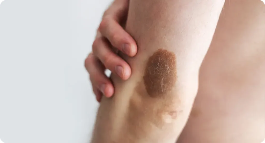
Congenital Melanocytic Naevi: What You Need to Know
Congenital melanocytic naevi (CMN) are pigmented birthmarks that appear at birth or shortly thereafter. They vary widely in size—from small spots to extensive patches covering large areas of skin. While small or medium CMNs typically pose minimal risk, larger or “giant” naevi (greater than 20cm in diameter in adulthood) carry a small but measurable risk of malignant transformation into melanoma.
This risk is heightened in early childhood and in individuals with multiple CMNs. Signs that a CMN needs evaluation include changes in size, colour variation, surface irregularities, ulceration, or the sudden appearance of nodules.
In addition to melanoma, giant CMNs may be associated with neurocutaneous melanosis—a rare condition involving excess pigment cells in the brain or spinal cord, which can lead to neurological symptoms. Dermatologists often monitor CMNs through photography, dermatoscopy, and, in some cases, biopsy or MRI. Early detection and routine assessment are crucial, especially if there’s a family history of skin cancer or if the naevus changes over time.
When Should You See a Dermatologist?

Not all birthmarks require medical intervention, but knowing when to see a dermatologist can prevent complications down the line. The general rule is: if a birthmark changes in size, colour, shape, texture, or begins to bleed or itch, it should be examined by a professional. Other red flags include rapid growth, irregular borders, or asymmetry—features similar to those seen in skin cancers. For children, it’s particularly important to assess whether a birthmark is interfering with function (e.g. vision or breathing) or development, especially if located near the eye, nose, or mouth.
Even birthmarks that appear benign may need review for aesthetic or psychological reasons. Some vascular birthmarks, especially port-wine stains on the face, can darken and thicken with age, potentially affecting confidence and social interaction. Dermatologists can advise on both medical and cosmetic considerations, using imaging and skin assessment tools to guide the best course of action. Early consultation also opens the door to modern treatment options, many of which are most effective when initiated in infancy or early childhood.
Laser Therapy: A Front-Line Treatment for Many Birthmarks
Laser therapy has emerged as a leading treatment for both vascular and pigmented birthmarks. Pulsed dye lasers (PDL) are commonly used to treat port-wine stains, working by targeting blood vessels in the dermis without damaging surrounding tissue. Results are best when treatment starts early, often in infancy, and multiple sessions may be needed for optimal results. For haemangiomas, laser treatment can complement oral medications like beta-blockers in shrinking or resolving the lesion.
In pigmented birthmarks such as café-au-lait spots or small CMNs, Q-switched lasers can break down excess melanin, gradually fading the lesion. However, outcomes can be variable and some pigmentation may return. Laser treatment is typically done under topical anaesthetic, with minimal downtime and a low risk of scarring. It’s essential that treatment is carried out by a qualified dermatologist or laser specialist with experience in paediatric dermatology when dealing with infants or toddlers. Regular follow-up ensures that results are maintained and any side effects are addressed promptly.
Surgical Removal: When Is It the Right Choice?
Surgical excision may be recommended for certain types of birthmarks, particularly when there’s a high risk of malignancy, functional interference, or significant cosmetic concern. This is most often the case with large or giant congenital melanocytic naevi, especially if located in high-friction areas or if changes suggest malignancy. Depending on the size and depth of the lesion, surgery may be completed in stages, and skin grafting or tissue expansion might be necessary to achieve a natural appearance post-removal.
In some cases, a combination of surgery and laser treatment offers the best outcome—removing the bulk of the lesion surgically, followed by laser resurfacing to improve skin texture or pigmentation. The decision to opt for surgery is typically made after multidisciplinary consultation, often involving dermatologists, plastic surgeons, and paediatric specialists.
Risks include infection, scarring, and the psychological impact of multiple procedures, which must be carefully weighed against the benefits. For families, clear communication with the clinical team and psychological support can make a major difference in navigating the process.
Psychological Considerations and Cosmetic Support

While the medical risks of birthmarks often take priority, the emotional and psychological impact—particularly for children and adolescents—should not be overlooked. Birthmarks in visible areas can lead to self-consciousness, bullying, or social anxiety, affecting a child’s self-esteem. Cosmetic camouflage products, such as specially formulated make-up or skin-coloured creams, can help individuals feel more confident while awaiting or recovering from treatment.
Psychodermatology—a growing field that addresses the psychological aspects of skin conditions—may also be beneficial. Support groups, counselling, or play therapy can help children process their experiences, particularly if the birthmark is large or requires repeated treatment. For adults, aesthetic dermatology offers a range of minimally invasive procedures to reduce discolouration and improve skin tone, such as chemical peels or microneedling, though these should be used cautiously on birthmarked skin and always under medical supervision.
Alternative Treatments and Emerging Therapies
While traditional methods like laser and surgery remain the mainstay of birthmark treatment, newer approaches are emerging. For example, propranolol—a beta-blocker traditionally used for heart conditions—has shown excellent efficacy in treating infantile haemangiomas when administered early. This medication works by shrinking the blood vessels, often leading to complete resolution without the need for surgery.
Research is also underway into gene-targeted therapies for vascular malformations and topical treatments that may reduce pigmentation in certain birthmarks. Stem cell therapy and regenerative medicine are at the experimental stage, with the potential to regenerate normal skin tissue in cases of large birthmark removal.
However, most of these options are still under clinical investigation and are not yet widely available outside of specialist centres. As the field progresses, the future may hold less invasive, more personalised treatments tailored to the specific cellular makeup of each birthmark.
Monitoring Birthmarks Over Time: What to Watch For
Birthmarks are dynamic, especially in early childhood, and their appearance may evolve over time. While many remain stable or fade, some can become more prominent or develop concerning features. Parents and individuals should regularly monitor birthmarks for any notable changes—especially in colour, size, texture, or symptoms such as pain or bleeding.
A useful guide is the “ABCDE” rule, often applied to moles: Asymmetry, Border irregularity, Colour variation, Diameter over 6mm, and Evolving appearance. If a birthmark demonstrates any of these features, particularly in pigmented lesions like congenital melanocytic naevi, prompt evaluation by a dermatologist is essential.
In addition, it’s vital to watch for new nodules within a pre-existing birthmark, sudden darkening, or any ulceration. Annual skin checks, either by a GP with dermatological training or a consultant dermatologist, can help ensure no changes are missed, particularly for those with a family history of melanoma or other skin disorders.
Birthmarks and Syndromic Conditions: When It’s More Than Skin Deep
Certain birthmarks are not just isolated skin features but may signal an underlying medical syndrome. For instance, large or multiple café-au-lait spots may be an early sign of neurofibromatosis type 1 (NF1), a genetic disorder that affects nerve tissue growth. Similarly, extensive port-wine stains affecting the face, particularly around the eye and forehead, may be linked to Sturge-Weber syndrome, which involves abnormal blood vessels in the brain and eye, leading to seizures or glaucoma.
Blue-grey Mongolian spots, though usually benign, when numerous or oddly located, can be mistaken for bruising and warrant careful evaluation. In these cases, dermatologists often work alongside paediatricians, neurologists, or geneticists to ensure comprehensive care. Early diagnosis of syndromic conditions allows for better management of both skin and systemic symptoms, and may improve long-term outcomes through targeted therapy or supportive intervention.
Laser Limitations and Considerations in Skin of Colour
While laser therapy is a highly effective treatment for many birthmarks, its success and safety can vary depending on the patient’s skin tone. Individuals with darker skin (Fitzpatrick skin types IV–VI) are at greater risk of complications such as post-inflammatory hyperpigmentation, hypopigmentation, and scarring following laser procedures.
As a result, specific laser types and lower energy settings are typically chosen to reduce these risks. Nd:YAG lasers, for example, penetrate more deeply and are often safer in pigmented skin. Moreover, treatment must always be carried out by professionals experienced in treating skin of colour, with pre- and post-care protocols tailored accordingly.
Unfortunately, people with darker skin tones have historically been underrepresented in dermatology studies, leading to a gap in evidence-based protocols. However, awareness is improving, and newer laser systems are being developed with greater precision and reduced risk, making it increasingly possible to offer safe and effective treatment across all ethnicities.
Birthmarks and Sun Protection: A Critical Preventative Step
Regardless of whether a birthmark is being treated or monitored, sun protection is a key element of skin care. UV exposure can darken pigmented birthmarks, increase irritation in vascular lesions, and in rare cases, accelerate malignant transformation. For children with large congenital melanocytic naevi or port-wine stains, broad-spectrum sunscreen with SPF 50+ should be used daily on exposed areas, even in winter. Physical barriers such as hats, UPF-rated clothing, and shaded prams offer additional defence.
It’s also advisable to avoid peak sun hours and reapply sunscreen every two hours when outdoors. Dermatologists often recommend mineral sunscreens for sensitive or lesion-prone skin, as these contain zinc oxide or titanium dioxide and are less likely to cause irritation. Encouraging sun-safe habits from an early age not only protects birthmarks but promotes overall skin health and reduces long-term cancer risk, especially in genetically predisposed individuals.
Choosing the Right Clinic or Specialist: What to Look For
When seeking treatment or evaluation for a birthmark, choosing the right clinician and clinic is crucial for both safety and results. Ideally, the first point of contact should be a consultant dermatologist with expertise in paediatric or medical dermatology, particularly when dealing with complex or atypical lesions.
For laser or surgical intervention, clinics should offer access to board-certified professionals with advanced training in dermatological lasers, paediatric sedation (if needed), and scar-minimising techniques. Multidisciplinary centres—often found in university hospitals or dedicated children’s hospitals—can provide coordinated care with input from dermatologists, plastic surgeons, radiologists, and psychologists.
Before proceeding with treatment, ask about the clinic’s success rates, before-and-after photos, and whether patch testing or trial laser pulses will be done. An ethical practitioner will be upfront about expected outcomes, number of sessions needed, potential complications, and realistic timelines for improvement. Transparency and tailored care should be the cornerstone of any reputable dermatology service.
Navigating Birthmark Care in the NHS and Private Sector
In the UK, birthmark assessment and treatment are available through both the NHS and private healthcare providers, though access and waiting times can vary. The NHS typically covers evaluation and treatment of birthmarks deemed medically necessary—such as those at risk of malignancy, causing functional impairment, or linked to syndromic conditions. For example, large congenital melanocytic naevi or haemangiomas affecting vision or breathing will usually be prioritised for referral to dermatology or specialist vascular anomaly clinics.
However, treatments considered cosmetic, like fading small café-au-lait spots or lightening port-wine stains for aesthetic reasons, may not be funded. In such cases, private care offers more immediate access to laser treatments and wider procedural options, often in combination with advanced imaging and follow-up.
It’s important for patients and parents to understand referral pathways: starting with a GP consultation, seeking a dermatologist’s opinion, and requesting referral to a tertiary centre if needed. For those opting for private care, ensure the practitioner is registered with the General Medical Council (GMC) and the clinic is CQC-regulated. Both sectors offer high standards of care, but informed navigation of the system ensures the right support is accessed at the right time.Bottom of Form
The Role of Genetic Testing and Dermatopathology in Complex Cases

In rare or complex birthmark cases, particularly those involving multiple lesions or suspected syndromic associations, genetic testing and dermatopathology can play a vital diagnostic role. Certain birthmarks—such as extensive café-au-lait spots, segmental capillary malformations, or large congenital melanocytic naevi—may be linked to underlying mutations that influence both skin and systemic health.
For instance, genetic analysis may confirm conditions like neurofibromatosis type 1 (NF1) or identify mosaic mutations in RAS genes associated with large CMN. In cases where malignancy is suspected, dermatopathologists analyse biopsy samples to assess cellular architecture, rule out melanoma, and guide treatment plans.
These specialists bridge the gap between clinical dermatology and molecular science, offering a deeper understanding of each lesion’s nature. While not every birthmark requires such detailed scrutiny, in selected patients this approach ensures more accurate diagnoses, personalised care, and improved outcomes—particularly when long-term surveillance or multidisciplinary management is needed.
Final Words
Most birthmarks are entirely harmless and require no intervention beyond routine observation. However, a small percentage carry medical or psychological risks that warrant professional assessment.
Knowing the type of birthmark, understanding warning signs, and seeking early dermatological advice can make all the difference in ensuring health, safety, and emotional wellbeing. Early evaluation is particularly important in children, where certain birthmarks may evolve over time or indicate underlying conditions.
Thanks to advances in laser technology, surgical methods, and pharmacological treatments, individuals today have more options than ever to manage and treat birthmarks effectively. Treatments are now more precise, less invasive, and tailored to the specific needs of each patient.
If you or your child has a birthmark you’re unsure about, don’t hesitate to consult a dermatologist—it’s a simple step that can provide reassurance or open the door to life-changing care. If you are looking for help in removing a birthmark, then you can contact us at The London Dermatology Centre for a consultation with one of our expert dermatologists who can guide you on the best approach.
References
- Bruckner, A.L. and Frieden, I.J., 2003. Hemangiomas of infancy. Journal of the American Academy of Dermatology, 48(4), pp.477–493.
- Haggstrom, A.N., Lammer, E.J., Schneider, R.A., Marcucio, R., Frieden, I.J. and the Hemangioma Investigator Group, 2006. Patterns of infantile hemangiomas: new clues to embryonic development. Pediatrics, 117(3), pp.698–703.
https://pediatrics.aappublications.org/content/117/3/698 - Tollefson, M.M. and Frieden, I.J., 2010. Early growth of infantile hemangiomas: what parents’ photographs tell us. Pediatrics, 127(6), pp.e1239–e1248.
- Léauté-Labrèze, C., Hoeger, P., Mazereeuw-Hautier, J., Guibaud, L., Baselga, E., Posiunas, G., Phillips, R.J., Glover, M., Harper, J.I., Bermudez, F., and Gelmetti, C., 2015. A randomized, controlled trial of oral propranolol in infantile hemangioma. New England Journal of Medicine, 372(8), pp.735–746.
https://www.nejm.org/doi/full/10.1056/nejmoa1404710 - Kinsler, V.A., O’Hare, P., Bulstrode, N. and Calonje, E., 2017. The management of congenital melanocytic naevi: best clinical practice. British Journal of Dermatology, 176(3), pp.539–551.
https://onlinelibrary.wiley.com/doi/full/10.1111/bjd.15012