


If you have ever been told that you need a skin biopsy, you may wonder why your dermatologist cannot tell what is wrong simply by looking at your skin. Sometimes a mole, persistent rash or unusual growth needs a closer look, and that is where dermatopathology becomes important.
Dermatopathology brings dermatology and pathology together, allowing a small sample of your skin to be examined under a microscope. Today, special stains and other laboratory tests can also be used when needed, helping your doctors understand changes that may not be visible from the surface.
Before Skin Biopsies: What Could Doctors See?
Before doctors could examine skin under a microscope, they had to rely largely on what they could see and feel. They looked at features such as colour, scaling and blisters, as well as symptoms such as itching and how the problem changed over time.
When microscopy developed in the nineteenth century, doctors could examine what was happening beneath your skin’s surface. This created the approach used today: comparing what can be seen on your skin with the microscopic changes found in a biopsy.
How Microscopy Revealed What Was Happening Beneath Your Skin
If your skin looks similar to several different conditions, examining it under a microscope can reveal details that are impossible to see during an ordinary skin examination. If you have a skin biopsy, it can reveal changes in your epidermis, dermis, hair follicles and glands that may not be visible during a clinical examination.
By comparing these microscopic findings with the appearance and history of your skin problem, doctors gained a more reliable way to distinguish between conditions. This connection between your clinical features and tissue findings became a foundation of modern dermatopathology.
How Did Tissue Stains Make Skin Changes Easier to See?
Tissue stains made it much easier for doctors to see cells and structures clearly under a microscope. The development of haematoxylin and eosin (H&E) staining in the nineteenth century created useful contrast between cell nuclei and surrounding tissue, helping pathologists recognise tissue structure and cellular patterns more clearly. By the mid-1870s, haematoxylin and eosin were being combined as histological stains, laying the foundations for the H&E method that remains central to pathology today.
When your biopsy is examined today, H&E is usually the basic stain used to reveal the structure of your skin and its cells. Depending on what your dermatologist is investigating, these findings may support a diagnosis or show that additional laboratory tests are needed.
Paul Gerson Unna and the Birth of Modern Dermatopathology
Paul Gerson Unna was an important figure in early dermatopathology, helping establish links between the clinical appearance of skin disease and its microscopic changes. His work helped define important microscopic changes such as acanthosis and spongiosis, terms you may still encounter in dermatopathology reports today.
Unna also improved the study of skin tissue through staining and microscopy. His approach helped establish the clinicopathological method of understanding skin disease by combining clinical appearance with microscopic findings.
Paul Gerson Unna’s Die Histopathologie der Hautkrankheiten, published in German in 1894 and translated into English in 1896 as The Histopathology of the Diseases of the Skin, became an important milestone in the development of dermatopathology. By systematically linking clinical skin disease with microscopic tissue changes, his work helped establish cutaneous histopathology as a more organised field of study.
The Skin Biopsy Turned Microscopy Into a Clinical Tool
A skin biopsy made it possible for you to have a small sample examined under a microscope, helping doctors understand what was happening beneath your skin. In 1887, Edward L. Keyes described the cutaneous punch, an early instrument that helped establish the use of punch techniques in dermatology and became a precursor to modern skin-biopsy punches.
Modern skin-biopsy techniques now allow your dermatologist to take a carefully selected sample for microscopic examination. The most appropriate method depends on what your doctor needs to investigate and how deep the relevant changes may be.
Why Are There Different Types of Skin Biopsy?
As skin biopsy became more established, doctors developed different ways to obtain tissue depending on what they needed to investigate. Punch biopsies provide a cylindrical sample that includes the epidermis and dermis and may extend into subcutaneous tissue, while shave techniques are useful for selected more superficial lesions. Incisional biopsy removes part of a lesion, whereas excisional biopsy removes the whole visible lesion when appropriate.
The technique matters because different diseases involve different depths and structures within the skin. A sample that is too superficial or taken from an unrepresentative area may not contain the changes needed for diagnosis. Modern biopsy planning therefore considers the suspected condition, lesion size, location and depth before the tissue is removed.
Why the Right Biopsy Site Matters for Your Diagnosis
When you have a skin biopsy, the location of the sample can be just as important as how it is examined. Your dermatologist needs to choose the right lesion and area because scratching, infection or healing can change what your tissue shows.
Biopsy planning therefore begins before your sample reaches the laboratory. Your dermatologist must select an appropriate site and technique so the dermatopathologist receives the most useful tissue for interpretation.
If you are having a skin biopsy, tell your dermatologist about creams, medicines or treatments you have recently used on the area and whether the lesion has been scratched, infected or changing. Choosing the right part of a lesion and providing relevant clinical history can help the dermatopathologist interpret the biopsy more accurately.
What Happens to Your Skin Biopsy Before It Is Examined?
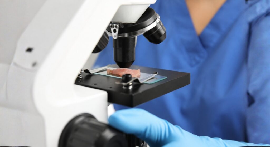
After a routine skin biopsy is taken, the tissue is usually fixed in formalin, processed, embedded in paraffin wax and cut into very thin sections with a microtome before being placed on slides and stained. Some specialised tests require different handling, so the laboratory method depends on what your dermatologist is investigating.
This careful preparation helps your dermatopathologist see important details clearly, allowing a tiny piece of your skin to be examined across several sections showing how your epidermis, dermis and other structures are changing.
What Is Your Dermatopathologist Looking for Under the Microscope?
When your biopsy is examined, your dermatopathologist does not simply look for one abnormal cell or feature. They look for patterns involving inflammation, changes in the epidermis, the position of blisters and other clues that can help narrow down the diagnosis.
In practice, your dermatopathologist can combine your clinical history with these microscopic patterns rather than simply matching the biopsy to a textbook image.
Why Your Clinical History Matters to Your Biopsy Result
A skin biopsy does not always give you the full answer on its own because different conditions can sometimes share similar microscopic features. Your dermatopathologist needs to know how the lesion looks, where it appears, how long you have had it and what symptoms you experience.
This approach is called clinicopathological correlation, where your clinical details are combined with your biopsy findings. Combining these findings allows your dermatologist and dermatopathologist to work together to reach the best-supported diagnosis.
In UK practice, the Royal College of Pathologists publishes a dedicated tissue pathway for dermatopathology covering the handling and reporting of many common dermatopathology specimens, including inflammatory dermatoses and specimens requiring immunofluorescence. Good dermatopathology depends not only on what is seen under the microscope but also on appropriate specimen handling and relevant clinical information supplied with the biopsy.
Why Specialist Dermatopathology Matters for Your Diagnosis
By the mid-twentieth century, dermatopathology had developed into a specialised field combining dermatology and pathology. Walter Lever’s Histopathology of the Skin, first published in 1949, and the establishment of the American Society of Dermatopathology in 1962 helped formalise dermatopathology as a recognised specialist field.
If your biopsy shows an unusual or complex microscopic pattern, specialist dermatopathology interpretation can be particularly valuable. Your dermatopathologist can compare the tissue findings with your clinical information and any additional test results to support the most appropriate diagnosis.
Key Milestones in Dermatopathology
| Period | Development | Why It Mattered |
| 19th century | Microscopy became increasingly important in medicine | Allowed doctors to examine skin below the visible surface |
| Mid-1870s | Foundations of H&E staining established | Made tissue structures easier to distinguish microscopically |
| 1887 | Edward L. Keyes described the cutaneous punch | Helped establish an early precursor of the modern punch-biopsy instrument |
| 1890s | Paul Gerson Unna advanced microscopic classification of skin disease | Strengthened links between clinical dermatology and pathology |
| 1949 | Walter Lever published Histopathology of the Skin | Became an influential reference in modern dermatopathology |
| 1962 | American Society of Dermatopathology established | Helped formalise the specialty |
| 1960s–1970s | Immunofluorescence became increasingly important | Improved diagnosis of autoimmune blistering diseases |
| Late 20th century | Immunohistochemistry expanded | Helped classify tumours and identify cellular proteins |
| Late 20th–21st century | Molecular testing developed | Added genetic information in selected difficult cases |
| Modern practice | Whole-slide imaging and AI tools developed | Support digital review, consultation, research and selected diagnostic workflows |
Why Might Your Biopsy Need Special Stains?
H&E staining provides your dermatopathologist with important information, but sometimes more detail is needed. Special stains can help your doctor identify fungi, bacteria, pigment, connective tissue and other features that may not be clear on routine staining.
This means your biopsy can be examined in different ways depending on what your dermatologist suspects. This can allow the same small tissue sample to provide more information about what your lesion contains and how its cells are behaving.
How Can Immunofluorescence Help Diagnose Blistering Diseases?
Direct immunofluorescence (DIF) helped doctors understand what was happening inside your skin during autoimmune blistering diseases. It can detect characteristic deposits of immunoglobulins and complement in the skin, helping specialists distinguish autoimmune blistering disorders such as pemphigus and pemphigoid.
This may mean having two biopsies taken during the same appointment. In suspected autoimmune blistering disease, one sample may be taken from a representative lesion for routine H&E microscopy, while another is often taken from carefully selected nearby skin for DIF and handled separately. This approach allows specialists to compare the structure of the skin on routine microscopy with characteristic immune deposits detected by DIF.
When Might Your Biopsy Need Immunohistochemistry?
Immunohistochemistry (IHC) uses antibodies to identify specific proteins within your skin cells. This can help your dermatopathologist determine what type of cells are present, especially when tumours look similar on routine H&E staining.
You may not need IHC if your biopsy is straightforward. When extra testing is needed, your dermatopathologist can combine the staining results with the H&E findings and your clinical information to support a more specific and well-evidenced diagnosis.
How Dermatopathology Helps Diagnose Skin Cancer
Dermatopathology plays an important role in helping your doctor diagnose and classify skin cancer. Your tissue sample can help determine whether a growth is benign or malignant and what type of tumour is present. Depending on the tumour and the type of specimen removed, the pathology report may also include features such as tumour depth or whether abnormal cells reach the surgical margins.
The pathology report can therefore help guide important decisions about further surgery or treatment. When your biopsy is difficult to interpret, additional tests such as immunohistochemistry or molecular testing may be used to provide a more reliable diagnosis.
What Your Dermatopathology Report May Tell You
A modern dermatopathology report can contain more than the name of a diagnosis. Depending on the condition and the type of specimen, your dermatopathologist may describe important microscopic findings, give a differential diagnosis when more than one condition is possible, or report features that help guide further management.
For some skin cancers, relevant information may include tumour type, depth or thickness and whether tumour reaches an examined surgical margin. The report may also include results from special stains, immunohistochemistry or molecular testing when these have been required. If the findings do not provide one definite answer, your dermatopathologist may recommend clinicopathological correlation, additional tests or another biopsy so that the result can be interpreted alongside your clinical picture.
When Might Molecular Testing Help Your Diagnosis?
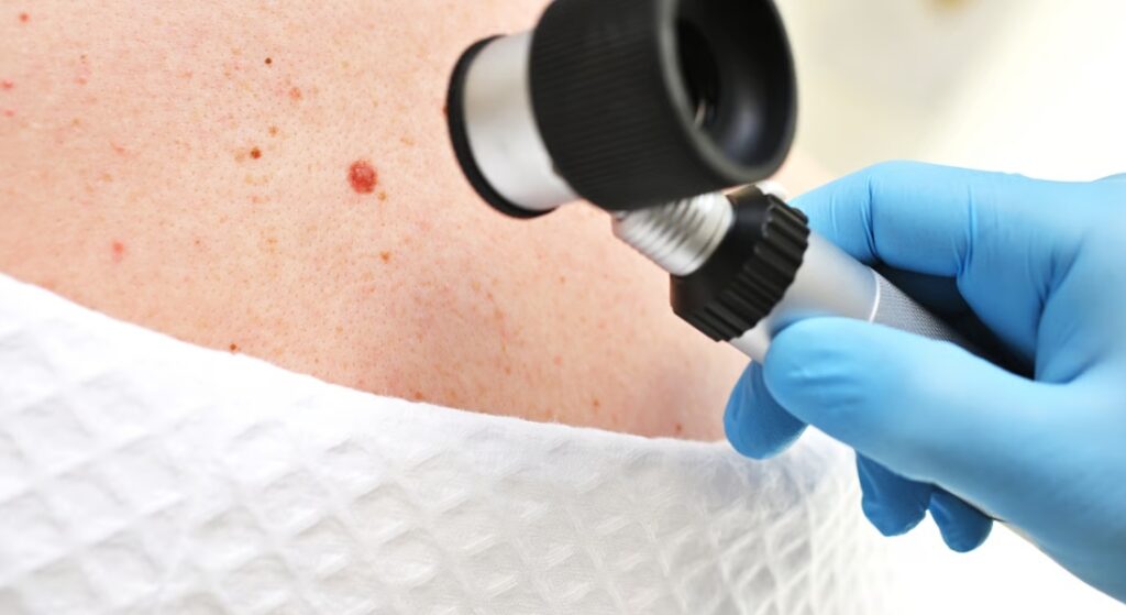
Most skin biopsies do not need genetic testing, but molecular tests can provide extra information in selected difficult cases. Techniques such as PCR, fluorescence in situ hybridisation and other molecular tests can help identify tumour-associated genetic alterations, inherited variants or microbial nucleic acid in selected cases.
When uncertainty remains, your specialist can interpret molecular findings alongside tissue appearance, immunohistochemistry and clinical information to help support the diagnosis.
How Digital Pathology May Support Your Biopsy Diagnosis
Digital pathology allows your biopsy slides to be scanned into highly detailed digital images that can be viewed, stored and shared on a computer. This can make it easier for your dermatopathologist to seek specialist opinions, teach trainees and compare difficult cases.
Artificial intelligence is also being explored to help recognise patterns and measure microscopic features. However, your diagnosis still depends on expert interpretation and clinical information, with digital tools supporting rather than replacing your dermatopathologist.
Myth vs Fact
| Myth | Fact |
| A skin biopsy always gives one definite diagnosis. | Not always. Some diseases share microscopic features, so clinical information and sometimes extra tests are needed. |
| Every skin biopsy is processed in exactly the same way. | No. Routine histology commonly uses formalin-fixed tissue, while tests such as direct immunofluorescence need different handling. |
| Dermatopathology is mainly used for skin cancer. | No. It is also important in inflammatory, infectious, autoimmune and inherited skin conditions. |
| The biopsy site does not matter as long as some skin is removed. | False. Choosing the correct lesion and area can be critical to obtaining useful diagnostic information. |
| Special stains and molecular tests are needed for every biopsy. | No. Many diagnoses can be made using routine H&E microscopy and clinical information alone. |
| Artificial intelligence now replaces dermatopathologists. | No. AI and digital tools can support selected tasks, but expert interpretation and clinical correlation remain essential. |
Frequently Asked Questions
1. What is dermatopathology and why might you need it?
Dermatopathology combines dermatology and pathology to examine your skin at a microscopic level. You may need it when a mole, rash, growth or other skin change cannot be diagnosed reliably from its appearance alone.
2. How did microscopy change the diagnosis of skin disease?
Before modern microscopy, doctors mainly relied on what they could see and feel on your skin. Microscopy allowed them to examine cells and tissue structures underneath the surface, making diagnosis more detailed and precise.
3. Why is a skin biopsy important for your diagnosis?
A skin biopsy gives your doctor a small sample of the affected skin for microscopic examination. This can help identify changes that cannot be seen from the surface and help distinguish between conditions that may look similar.
4. How does your dermatologist choose the right biopsy site?
The location and type of biopsy can affect the accuracy of your diagnosis. Your dermatologist will choose a representative area while considering the appearance, age and location of the lesion and avoiding areas that may have been significantly altered by scratching or treatment.
5. What happens to your skin biopsy in the laboratory?
For routine histology, your biopsy is usually fixed, processed and embedded in paraffin wax before being cut into very thin sections. The sections are placed on slides and stained for microscopic examination. Some specialised tests, including direct immunofluorescence, require different specimen handling.
6. What is H&E staining and why is it used?
Haematoxylin and eosin, or H&E, is a basic stain used to show the structure and arrangement of cells and tissues in your biopsy. It remains one of the most important starting points for diagnosing many skin conditions.
7. What are special stains used for in dermatopathology?
Special stains can help your dermatopathologist identify features such as fungi, bacteria, pigment or particular tissue components. Your biopsy may receive additional stains when routine H&E examination does not provide enough information.
8. How can immunohistochemistry help diagnose your skin condition?
Immunohistochemistry uses antibodies to identify specific proteins within cells. If your biopsy contains a tumour or another difficult-to-classify lesion, these markers can help your dermatopathologist determine which type of cells are present.
9. Can dermatopathology help diagnose skin cancer?
Yes. Your biopsy can help determine whether a growth is benign or malignant and identify the specific type of skin cancer. Additional tests, including immunohistochemistry or molecular testing, may be used when the diagnosis is difficult.
10. How is modern dermatopathology different from early skin diagnosis?
Modern dermatopathology combines your clinical history and examination with microscopy, special stains, immunohistochemistry and, when needed, molecular testing. Digital pathology and artificial intelligence are also being developed to support specialists, although expert interpretation remains essential.
Final Thoughts: What Dermatopathology Means for Your Diagnosis
If you have a skin biopsy today, there is far more happening behind the scenes than simply looking at a sample under a microscope. What began with doctors examining visible skin changes has developed into a specialist field that can combine microscopy with special stains, immunohistochemistry and, when needed, molecular testing.
For you, the important point is that a biopsy result is rarely interpreted in isolation. Your dermatologist and dermatopathologist can combine what is seen in your skin with your medical history and the laboratory findings, helping them reach the best-supported diagnosis and decide what should happen next.
References:
- Bhawan, J. (2006) ‘The evolution of dermatopathology—the American experience’, The American Journal of Dermatopathology, 28(1), pp. 67–71. Available at: https://pubmed.ncbi.nlm.nih.gov/16456331/
- Nguyen, R., Persaud, N.A. and Franco Garcia, A. (2025) ‘Paul Gerson Unna: A medical pioneer in dermatopathology’, Cureus, 17(5), article e84288. Available at: https://pmc.ncbi.nlm.nih.gov/articles/PMC12169496/
- Sina, B., Kao, G.F., Deng, A.C. and Gaspari, A.A. (2009) ‘Skin biopsy for inflammatory and common neoplastic skin diseases: Optimum time, best location and preferred techniques. A critical review’, Journal of Cutaneous Pathology, 36(5), pp. 505–510. Available at: https://pubmed.ncbi.nlm.nih.gov/19187117/
- Ferringer, T. (2015) ‘Immunohistochemistry in dermatopathology’, Archives of Pathology & Laboratory Medicine, 139(1), pp. 83–105. Available at: https://pubmed.ncbi.nlm.nih.gov/25549146/
- Braun-Falco, M. (2009) ‘Molecular diagnosis in dermatopathology: What makes sense, and what doesn’t’, Experimental Dermatology, 18(1), pp. 12–23. Available at: https://pubmed.ncbi.nlm.nih.gov/19054055/
- Royal College of Pathologists (2024) Tissue pathways for dermatopathology. G075, Version 3. Available at: https://www.rcpath.org/resourceLibrary/g075-tissue-pathways-for-dermatopathology.html