")

Moles, also known as naevi, are small clusters of pigmented cells that appear on the skin. Most people have them, and in the majority of cases, they are completely harmless. They can be present from birth or develop over time, often appearing during childhood and early adulthood. Many moles remain unchanged for years, and some may even fade away with age.
However, not all changes in moles are harmless. Paying attention to alterations in their shape, size, or colour is important because these can sometimes be early warning signs of skin cancer, including melanoma the most serious type. Early detection plays a crucial role in successful treatment, so knowing what to look for is essential.
A mole that suddenly grows larger, develops an irregular outline, changes in colour, becomes itchy, or starts to bleed should never be ignored. Even if you’ve had the mole for many years without any issues, new developments could indicate that something is wrong. Skin cancer can develop anywhere on the body, even in areas not regularly exposed to the sun, so a full-body awareness approach is key.
In this article, we’ll walk you through the ABCDE guide a simple method for identifying potentially dangerous changes in moles as well as when it’s time to book an appointment for a professional mole check. We’ll also explain what typically happens during a dermatologist-led mole assessment, so you know exactly what to expect and can feel confident about taking the right steps for your skin health.
What to Look for: The ABCDE Guide
The ABCDE guide is a widely recommended method for checking moles at home. It’s simple enough to remember but powerful in helping spot possible early signs of skin cancer particularly melanoma. By regularly using this checklist, you can become more aware of changes in your skin and seek medical advice before a problem develops into something more serious.
If you notice any of the following features in a mole, or in a new spot that has recently appeared, it’s time to arrange a skin check with a qualified dermatologist.
A – Asymmetry
A healthy mole is generally symmetrical. Imagine drawing an invisible line through the middle in a harmless mole, both halves will look roughly the same in size, shape, and colour distribution.
In contrast, a suspicious mole may be asymmetrical, meaning one half looks noticeably different from the other. This imbalance could be subtle, such as one side being darker, more raised, or oddly shaped. Because melanoma cells grow unevenly, this lack of symmetry is often one of the earliest warning signs.
B – Borders
Normal moles have smooth, well-defined borders where the pigment blends seamlessly into the surrounding skin.
If the edges are irregular, jagged, blurred, or have a notched appearance, it’s worth getting them assessed. Border irregularity occurs because cancerous cells grow in an uncontrolled, uneven way, creating an outline that doesn’t look neat or consistent. Sometimes the borders may appear to fade in and out or look smudged against your skin.
C – Colour
Most harmless moles have one consistent colour, usually a uniform shade of light to dark brown, tan, or pink, depending on your skin tone.
Be alert if your mole contains multiple colours or an uneven mix of shades. A single mole showing black, deep blue, reddish, or even white areas can be a sign that different types of pigment are being produced by abnormal cells. Uneven colouring is particularly important to check if it appears alongside other changes, such as a growing size or irregular shape.
D – Diameter
Benign moles are often small typically less than 6mm in diameter (about the width of a pencil eraser).
If your mole is larger than this, especially if it wasn’t always that size, it’s worth keeping a close eye on it. That said, melanoma can still occur in smaller moles, so diameter should never be the only factor you assess. Think of it as one piece of the puzzle rather than the whole picture.
E – Evolving
Perhaps the most important sign to watch for is change over time. A mole that evolves whether it grows larger, alters its shape, darkens, lightens, becomes raised, or develops new symptoms such as itching, bleeding, or crusting warrants medical attention.
Healthy moles tend to remain stable for years. If you notice one behaving differently, don’t wait and see if it settles down book a dermatology appointment. Early detection of melanoma dramatically improves treatment outcomes.
When to Get a Mole Checked by a Dermatologist
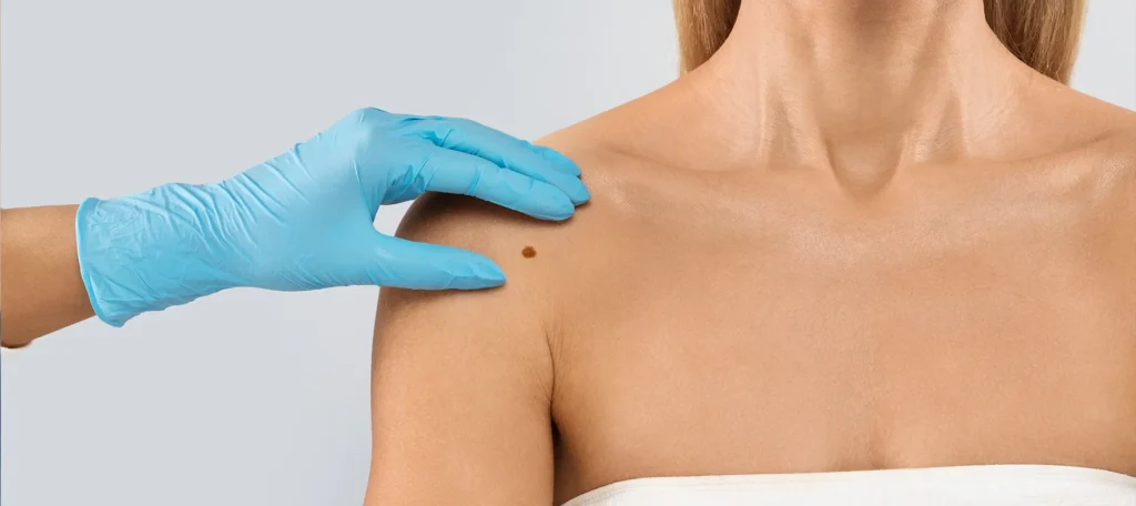
Even though most moles are harmless, it’s always better to be cautious when it comes to changes in your skin. The earlier a potential problem is identified, the easier it is to treat. In fact, melanoma detected at an early stage has a far higher survival rate than cases found later, making prompt attention essential.
If you notice one or more of the ABCDE characteristics we discussed earlier or any other unusual change book an appointment with a dermatologist as soon as possible. A dermatologist is trained to distinguish between harmless moles and those that require further investigation, often using specialised tools such as a dermatoscope for a closer look at skin structures invisible to the naked eye.
You should also be aware that changes can occur even in moles you’ve had for years, so don’t assume an “old” mole is safe just because it has been there a long time. Skin cancer can develop anywhere, including areas that are not often exposed to sunlight, such as the soles of the feet, the scalp, or under the nails.
Signs that it’s time to book a mole check include:
- Changes in shape, size, or colour – especially if the mole becomes irregular, darker, lighter, or develops multiple shades.
- Persistent symptoms – such as pain, tenderness, itching, or burning sensations in or around the mole.
- Bleeding, oozing, or crusting – even minor surface changes can be a warning sign.
- New and unusual moles – particularly those that look different from the rest of your moles (this is called the “ugly duckling” sign).
- Rapid growth – a mole that seems to be getting bigger quickly should never be ignored.
- Moles in hard-to-see places – such as the back, scalp, or behind the ears. These should be checked regularly by someone you trust or during a professional skin exam.
It’s worth remembering that not every mole showing these changes will turn out to be cancerous. However, the only way to be sure is through a professional assessment. Dermatologists can often provide reassurance on the spot, or, if there’s any uncertainty, arrange for a biopsy to examine the tissue under a microscope.
Regular mole checks both at home and with a professional are a simple but powerful step in protecting your skin health.
What to Expect During a Mole Check

If you’ve booked a mole check, you might feel a little anxious about what will happen during the appointment especially if it’s your first time. Knowing the process in advance can help you feel more at ease and prepared. Dermatologists carry out mole assessments regularly, and the procedure is straightforward, thorough, and designed to give you peace of mind.
Here’s what typically happens during a mole check:
1. Skin Examination
Your appointment will usually begin with a discussion about your concerns, medical history, and any changes you’ve noticed in your moles. The dermatologist will then conduct a full-body skin examination to assess both the mole in question and the rest of your skin.
This includes checking areas that are often overlooked, such as:
- The scalp and hairline
- Behind the ears
- The back and buttocks
- Under the breasts
- The soles of your feet and between the toes
- Under and around the nails
The reason for such a thorough check is that skin cancer can appear anywhere, not just in sun-exposed areas. The dermatologist will use the ABCDE guide to evaluate each mole and note any that require closer inspection.
2. Dermoscopy
In many cases, the dermatologist will use a dermoscope a handheld device that magnifies the skin and uses polarised light to reveal details invisible to the naked eye.
Dermoscopy allows the dermatologist to examine:
- Pigment patterns
- Blood vessel structures
- Subtle changes in colour and texture
These details help them distinguish between harmless moles and ones that might need further testing. Dermoscopy is quick, completely painless, and does not require any preparation on your part.
3. Biopsy (If Needed)
If a mole appears suspicious, the dermatologist may recommend a biopsy. This involves removing either a small section or the entire mole so that the cells can be examined under a microscope.
Key points to know about a biopsy:
- It’s usually done under local anaesthetic, so you won’t feel pain during the procedure.
- The process is quick often completed within 15–30 minutes.
- Recovery is minimal, with aftercare instructions provided to help the area heal.
The biopsy results will confirm whether the mole is benign (harmless) or cancerous, allowing your dermatologist to decide the next steps.
4. Follow-Up Recommendations
Once the examination is complete, your dermatologist will explain the findings and discuss any necessary action.
- If the mole is benign, you’ll be advised on self-monitoring techniques and the frequency of future skin checks.
- If the mole is cancerous or pre-cancerous, your dermatologist will talk you through treatment options, which might include surgical removal, cryotherapy, or other therapies depending on the type and stage of the cancer.
Even if no immediate action is needed, your dermatologist may recommend routine skin checks especially if you have many moles, a family history of skin cancer, or fair skin that burns easily.
Why Skin Cancer Early Detection is Crucial
Skin cancer is not only one of the most common cancers worldwide, but also one of the most preventable and treatable provided it’s detected early. Each year, thousands of people are diagnosed with skin cancers ranging from the more common basal cell carcinoma (BCC) and squamous cell carcinoma (SCC) to the more aggressive and potentially life-threatening melanoma.
While BCC and SCC tend to grow slowly and are less likely to spread, melanoma behaves differently. It can develop quickly and, if left untreated, spread to other organs such as the lungs, liver, or brain. This process, known as metastasis, can make treatment far more challenging and significantly reduce survival rates.
The power of early detection cannot be overstated. When melanoma is identified at an early stage before it has penetrated deeply into the skin or reached the lymphatic system the five-year survival rate can be as high as 98%. At this point, treatment may involve nothing more than a minor surgical procedure to remove the mole and a small margin of surrounding tissue. In many cases, patients can return to normal life within days.
However, if melanoma is diagnosed once it has spread to distant parts of the body, survival rates drop sharply, sometimes to as low as 15–20% depending on the stage and location of spread. At this point, treatment often requires more complex and prolonged approaches, such as:
- Immunotherapy, which stimulates the body’s own immune system to fight cancer cells.
- Targeted therapies, which block specific genetic mutations in the cancer.
- Radiotherapy or chemotherapy in certain cases.
While these treatments have improved outcomes in recent years, they can still be physically and emotionally demanding, with side effects that impact daily life.
Why timing matters so much
The skin is unique among organs because it’s visible. Unlike cancers that develop deep inside the body where symptoms may appear only in advanced stages skin cancer often gives us visible warning signs. A new mole, a change in an existing mole, or an unusual patch of skin is an opportunity to act early. Yet, despite these clear signals, many people delay getting checked, assuming it’s “probably nothing” or waiting to see if it goes away on its own.
Unfortunately, skin cancer rarely resolves without intervention. The longer you wait, the more chance abnormal cells have to multiply and spread. This is why dermatologists stress the importance of regular self-examination and professional skin checks. For most people, a monthly self-check combined with an annual or biannual dermatologist appointment can dramatically reduce the risk of late-stage diagnosis.
Who should be extra vigilant?
While everyone should monitor their moles, some groups face a higher risk of developing skin cancer and should be particularly proactive:
- People with fair skin, light hair, and light eyes, who tend to burn easily.
- Individuals with a history of frequent sunburns, especially during childhood.
- Those who spend a lot of time outdoors for work or recreation without consistent sun protection.
- People with a large number of moles or atypical (dysplastic) moles.
- Anyone with a family history of melanoma or other skin cancers.
- People with a weakened immune system due to illness or medication.
For these groups, dermatologists may recommend more frequent professional checks sometimes every 3–6 months in addition to self-monitoring.
The bottom line
Catching skin cancer early can mean the difference between a small, quick procedure and a lengthy battle with advanced disease. It’s one of the few cancers where you can spot the warning signs with your own eyes and act before it spreads. Taking a few minutes each month to examine your skin could quite literally save your life.
Final Thought: Prioritise Your Skin Health with Timely Checks
If you notice any changes in a mole or develop new moles that seem unusual, it’s important to take action and get them checked by a dermatologist. Early detection of skin cancer can make all the difference, so don’t hesitate to seek professional advice.
You can get in touch with us to book a consultation with our mole check clinic in London. Our expert dermatologists can assess any concerns you may have and guide you on the next steps for treatment or monitoring.
References:
- American Academy of Dermatology. (n.d.) What to look for: ABCDEs of melanoma. Available at: https://www.aad.org/public/diseases/skin-cancer/find/at-risk/abcdes
- Ebell, M. H. (2008) ‘Clinical diagnosis of melanoma’, American Family Physician, 78(10), pp. 1205–1208. Available at: https://www.aafp.org/pubs/afp/issues/2008/1115/p1205.html
- Vestergaard, M. E., Macaskill, P., Holt, P. E. and Menzies, S. W. (2008) ‘Dermoscopy compared with naked eye examination for the diagnosis of primary melanoma: a meta-analysis of studies performed in a clinical setting’, British Journal of Dermatology, 159(3), pp. https://www.aafp.org/pubs/afp/issues/2008/1115/p1205.html?utm_source=chatgpt.com
- MDPI. (2023) ‘Advances in early detection of melanoma and the future of …’, Life [online]. Available at: https://www.mdpi.com/2075-1729/13/4/974
- Nanci (PMC). (2011) ‘Novice Identification of Melanoma: Not Quite as Straightforward as …’, Acta Dermato-Venereologica. Available at: https://pmc.ncbi.nlm.nih.gov/articles/PMC3325479