


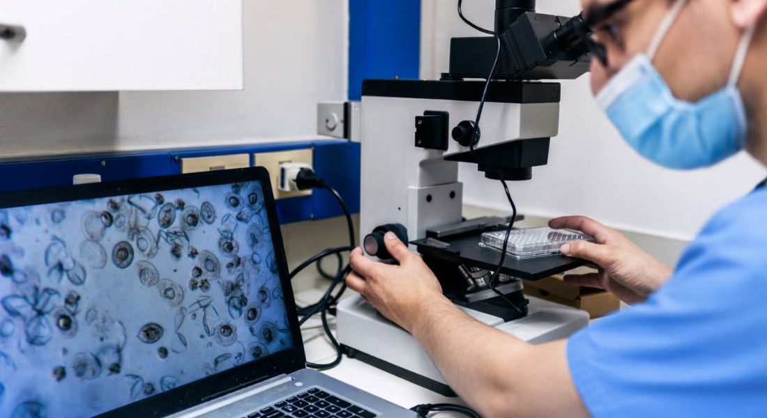
When you visit a dermatology clinic, most diagnoses begin with visual examination. Experienced clinicians can detect subtle colour, texture, and structural changes in the skin. However, even the most skilled eye relies on what is visible at the surface.
Molecular imaging introduces a different dimension. Instead of only observing structure, this technology explores biological activity beneath the skin. It aims to detect cellular and molecular changes before they become clinically obvious.
In 2026, research into molecular imaging is expanding rapidly. Scientists are exploring how advanced scanning techniques may reveal inflammation, immune activation, and early malignancy sooner than traditional methods. This article explains what molecular imaging involves and how it could shape the future of dermatology.
What Is Molecular Imaging?
Molecular imaging is a way of looking beneath the surface of what’s happening in your body right down at a cellular and biochemical level. Instead of simply showing the shape or structure of tissue, it detects specific molecular signals. In other words, it helps clinicians see how your tissue is behaving, not just what it looks like.
Traditional imaging, like standard scans, mainly shows structure whether something is thicker, larger, or physically damaged. Molecular imaging goes a step further by revealing function. It can highlight biochemical activity and cellular changes before any obvious structural damage appears. That means potential problems may be spotted earlier, sometimes before you’d notice visible symptoms.
In dermatology, this could mean identifying inflammatory pathways or early malignant cell changes much sooner. For you, that translates into the possibility of earlier intervention and more targeted treatment. It’s a shift from reacting to visible disease towards anticipating it and that has genuinely transformative potential.
Why Early Detection Matters in Dermatology
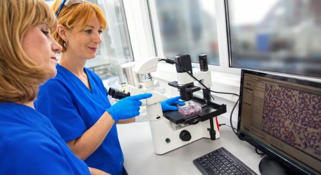
Many skin conditions begin quietly, at a microscopic level. Inflammatory cascades can start well before you see redness, swelling or scaling on the surface. In the same way, malignant cells may begin multiplying long before a mole changes shape or colour. By the time something is visible, the underlying process has often been active for some time.
Earlier detection gives you a valuable head start. If inflammation or abnormal cell activity is identified sooner, treatment can begin earlier too. That can slow progression, reduce severity and, in some cases, significantly improve long-term outcomes. Acting promptly often limits the amount of tissue damage that develops.
When care depends solely on visible signs, there’s an unavoidable delay. You might only seek help once symptoms become obvious or uncomfortable. Molecular imaging aims to narrow that gap by identifying biological activity before it becomes clinically apparent.
For you, that means a more proactive approach to diagnosis. Instead of waiting for disease to declare itself, clinicians can respond to early signals. Precision improves when care is guided by what’s happening beneath the surface, not just what can be seen.
Limitations of Visual Examination Alone
Visual examination is still the backbone of dermatology. An experienced clinician can learn a great deal just by looking carefully at your skin. However, some conditions are extremely subtle in their early stages. Pigmented lesions, in particular, can be difficult to interpret when changes are minimal or ambiguous.
Dermatoscopy certainly improves assessment. By magnifying and illuminating the skin, it helps reveal structural patterns that aren’t visible to the naked eye. Even so, it remains focused on morphology what the lesion looks like. It doesn’t directly measure what’s happening at a cellular or biochemical level beneath the surface.
This is where molecular imaging has the potential to complement existing tools. By adding functional insight to structural assessment, it may provide a fuller picture of what’s truly going on in your skin. Combining visual expertise with molecular data could enhance diagnostic accuracy, particularly in challenging or early-stage cases.
Imaging Inflammatory Activity
When you look at your skin, you’re only seeing part of the story. Inflammatory skin diseases involve immune activity happening beneath the surface often well before redness or swelling becomes visible. That’s where advanced molecular imaging becomes interesting.
1. Detecting what you can’t yet see: Molecular imaging techniques aim to identify inflammatory markers in the skin before symptoms appear. Instead of waiting for visible redness, clinicians could potentially detect immune activation at a microscopic or biochemical level. For you, that means insight before escalation.
3. Early cytokine activity as a warning sign: In many inflammatory conditions, cytokines signalling proteins involved in immune response increase before a flare becomes obvious. If imaging can detect rising cytokine activity early, it may allow pre-emptive treatment adjustments. That shift turns reactive care into proactive care.
4. Redness is only the surface expression: Visual redness is simply one outward sign of a deeper biological process. By the time you see flushing, scaling, or swelling, immune signalling has often been active for some time. Molecular signals typically precede what’s visible.
5. Moving towards predictive dermatology: If inflammatory activity can be monitored beneath the surface, management becomes more strategic. Instead of responding after symptoms develop, clinicians could intervene at the earliest measurable change.
For you, the significance lies in timing. The earlier inflammation is identified, the more controlled and measured your treatment adjustments can be. Imaging inflammatory activity brings dermatology closer to predictive, personalised care guided by biology rather than appearance alone.
Optical Coherence Tomography Advances
Optical coherence tomography, or OCT, creates detailed cross-sectional images of your skin almost like an optical ultrasound. It allows clinicians to see beneath the surface without making an incision. More advanced versions can now detect subtle microstructural and even vascular changes, which helps with analysing suspicious lesions in greater depth.
Newer systems are pushing this further by attempting to integrate functional information alongside structural imaging. In other words, it’s not just about what the tissue looks like, but how it’s behaving. Combining high-resolution structure with emerging molecular markers increases diagnostic precision, and the technology is continuing to evolve rapidly.
For you, this could mean earlier and more confident differentiation between benign and malignant lesions. The clearer the information, the more assured clinical decision-making becomes. Greater accuracy doesn’t just guide treatment it also provides reassurance when results are favourable.
Fluorescence-Based Imaging
Fluorescence-based imaging works by using light-reactive markers that attach to specific molecules in your skin. When exposed to certain wavelengths of light, these markers glow in distinctive patterns. Because abnormal or diseased cells often process or accumulate molecules differently from healthy cells, they can light up in a way that draws attention to areas of concern.
In skin cancer research, these fluorescence patterns may help identify early malignant changes before anything obvious appears on the surface. So even if a lesion looks fairly ordinary to you or even under standard examination subtle biochemical differences might already be detectable. Catching those changes earlier can significantly improve prognosis and guide more timely treatment.
It’s important to remember that this technology is still being refined. Researchers are working to improve accuracy, consistency and practical usability in everyday clinics. Clinical integration requires robust validation to ensure results are reliable and reproducible.
That said, early findings are encouraging. If ongoing studies continue to show strong predictive value, fluorescence imaging could become a valuable addition to dermatology. For you, that would mean another layer of precision supporting earlier, more confident decision-making.
Molecular Imaging in Skin Cancer
Early melanoma detection is absolutely critical. The earlier malignant change is identified ideally before deeper invasion occurs the better the survival rates. Molecular imaging has the potential to strengthen this window of opportunity by highlighting abnormal cellular behaviour before obvious structural changes are visible on your skin.
Some advanced scanners aim to detect altered metabolic activity, as cancer cells often use energy differently from healthy cells. By imaging these metabolic patterns, clinicians gain an additional layer of diagnostic information. When combined with dermoscopy and, where needed, biopsy, molecular imaging could refine diagnostic precision. Earlier, more confident intervention doesn’t just improve outcomes it can quite literally save lives.
Role in Psoriasis Monitoring
Psoriasis is driven by immune-mediated inflammation, even when the surface changes are what you notice first. Molecular imaging may one day help detect cytokine expression and other inflammatory signals beneath visible plaques. That would give clinicians a more objective measure of how active the disease really is, rather than relying only on what can be seen.
Sometimes plaques look relatively calm, but subclinical inflammation is still simmering underneath. If imaging could identify that hidden activity, it might explain why flares recur or why symptoms return quickly after treatment is reduced. Spotting that invisible inflammation could guide earlier or more targeted adjustments.
For you, this supports a more personalised approach to care. Therapy might be intensified when molecular activity rises or reduced when inflammation is genuinely quiet. Instead of static treatment plans, monitoring becomes dynamic responding to what’s actually happening beneath the surface of your skin.
Application in Eczema
Eczema flares usually begin with barrier breakdown and immune activation beneath the surface of your skin. Long before you notice obvious redness or itching, inflammatory signalling may already be underway. Molecular imaging has the potential to detect these early changes, giving you an indication that a flare could be developing.
If microvascular shifts or cytokine activity are identified early, you and your clinician might act sooner increasing moisturising, adjusting topical steroids, or introducing anti-inflammatory treatment before symptoms escalate. That kind of pre-emptive approach could reduce flare severity and shorten recovery time. Instead of constantly reacting, you’d be staying one step ahead.
Research in this area is still ongoing, and broader clinical adoption depends on strong validation. The technology needs to prove that it’s accurate, reliable and practical in everyday settings. Even so, the concept is compelling especially if it helps you move from flare management to genuine flare prevention.
Imaging Pigmentary Disorders
Pigmentary disorders often stem from melanocyte dysfunction the cells responsible for producing melanin in your skin. Long before you notice visible lightening or darkening, subtle changes in melanin distribution may already be occurring. Molecular imaging has the potential to detect these early alterations, which could help guide more targeted and timely treatment decisions.
In conditions such as vitiligo, immune-mediated destruction of melanocytes happens beneath the surface before depigmentation becomes fully apparent. If this activity could be identified early, there may be a window to intervene and potentially preserve pigmentation. Timing can make a meaningful difference in how well treatments work.
Being able to visualise these processes objectively would shift care from reactive to proactive. Instead of waiting for patches to expand, clinicians could respond to underlying immune activity as it begins. That may improve stability and reduce progression.
Precision imaging also allows pigment stability to be monitored more accurately over time. Rather than relying solely on visual comparison, subtle molecular changes could be tracked. For you, that means more personalised, nuanced care tailored to how your skin is actually behaving.
Precision Treatment Planning

Molecular imaging fits naturally into the move towards precision medicine. Instead of basing your treatment purely on standard protocols or average response rates, decisions could reflect your individual biological activity. That means therapy becomes more tailored to what’s actually happening in your skin, rather than what typically happens in most people.
Rather than automatically escalating or maintaining treatment at a fixed intensity, clinicians may one day adjust therapy according to molecular markers. That helps reduce overtreatment while also avoiding undertreatment. When biological feedback guides care, safety improves and strategies feel more individualised and that often translates into greater confidence and satisfaction with your treatment plan.
Potential Impact on Biologic Therapy
If you’re being considered for biologic treatment, the biggest question is always: is this the right pathway to target for you? Biologic medications are designed to block very specific immune signals. Choosing correctly from the outset makes a huge difference.
1. Confirming pathway activation before prescribing: Molecular imaging could potentially show which inflammatory pathway is active in your skin before treatment begins. Instead of relying purely on clinical patterns, your clinician could confirm that the pathway a biologic targets is actually driving your disease. That means more precise selection from the start.
2. More accurate response monitoring: Visible improvement can take time. However, a reduction in inflammatory markers may occur earlier at a molecular level. If imaging shows that immune signalling is decreasing, you and your clinician gain reassurance that the treatment is working even before you see full surface change.
3. Objective guidance for continuation or adjustment: If inflammatory markers remain elevated despite treatment, that may suggest the pathway isn’t being adequately controlled. Objective data helps guide whether to continue, optimise, or switch therapy, reducing prolonged trial-and-error cycles.
4. Improved cost-effectiveness: Biologics are significant investments, both personally and within healthcare systems. More accurate targeting reduces unnecessary switching and shortens ineffective treatment periods. For you, that means faster control and potentially fewer months of uncontrolled inflammation.
Ultimately, molecular imaging could make biologic therapy more strategic. Instead of prescribing based largely on patterns and probabilities, treatment decisions could be aligned with measurable immune activity improving precision, efficiency, and confidence in your care plan.
Challenges in Clinical Integration
Molecular imaging is still a costly technology, which means access isn’t yet widespread. For you, this could mean that only certain clinics or research centres offer these advanced assessments. On top of that, clinicians need specialised training to interpret the results accurately, so not every practitioner is equipped yet.
Standardisation is another hurdle. Different imaging systems might give slightly different readings, so results aren’t always directly comparable. For you, this means consistency can vary depending on where you go and which technology is used.
Clinical guidelines for incorporating molecular imaging into everyday dermatology are still evolving. Widespread adoption will depend on strong evidence showing clear benefits. Until then, careful, selective implementation is key, ensuring that any use of the technology genuinely supports your care.
Ethical and Data Considerations
Advanced molecular imaging captures a lot of detailed biological information, which makes protecting your confidentiality really important. Knowing that your data is handled ethically helps build trust, so you can feel confident that it’s being used responsibly.
It’s also essential that you understand why the imaging is being done and what it could reveal. Informed consent isn’t just a formality it ensures you know what’s happening with your data. Clear communication and strong regulation mean that innovation in this field can progress safely while keeping your interests front and centre.
Will Molecular Imaging Replace Biopsy?
A biopsy is still the gold standard when it comes to getting a definitive diagnosis. For you, this means that molecular imaging isn’t likely to replace it completely, but it can help reduce the number of unnecessary biopsies.
Molecular imaging can spot suspicious areas earlier than the naked eye might. It can also guide your clinician to the most appropriate spot for a biopsy, making the procedure more precise and effective.
Using imaging alongside traditional biopsy can make the whole process more efficient. You benefit because interventions are better informed, while tissue confirmation still ensures accuracy and safety.
Impact on Early Skin Cancer Screening
In the future, skin cancer screening programmes might start using advanced molecular imaging more widely. For you, this could mean earlier detection, especially if you’re considered high-risk. Monitoring becomes more sensitive, picking up changes before they’re visible.
Regular scans could reveal subtle malignant changes that might otherwise go unnoticed. Catching lesions at an early stage makes them far easier to treat, and your chances of a positive outcome improve significantly.
Wider adoption of these technologies depends on accessibility. Equipment costs and practical considerations will influence how quickly it’s rolled out.
Ultimately, evidence from ongoing research will determine the scale of implementation. For you, this means that while the technology is promising, it’s being carefully evaluated to ensure it truly benefits patients.
The Role of Artificial Intelligence
Artificial intelligence can make a real difference in how molecular imaging is interpreted. For you, this means that algorithms can spot patterns that might be too subtle for the human eye, improving the accuracy of early detection.
AI also allows large amounts of imaging data to be analysed quickly. Subtle molecular changes may be recognised sooner, giving your clinician more precise information to guide treatment decisions.
It’s important to remember that AI supports rather than replaces your doctor. Your clinician’s expertise remains central, and the best results come from combining human judgement with smart technology.
Limitations and Current Evidence
If you’re excited by new molecular imaging technologies, it’s important to remember that most of them are still in the research stage. Large-scale clinical trials are underway, but widespread clinical use depends on thorough validation.
1. Still mostly experimental: Many imaging techniques show promise in small studies or laboratory settings, but they haven’t yet been tested in large, diverse patient populations. For you, this means access may be limited to clinical trials for now.
2. Evidence must prove reliability: Not every innovation that looks impressive in early research translates into routine practice. Consistent accuracy, reproducibility, and meaningful clinical benefit are all required before a technology becomes standard care.
3. Careful evaluation continues: Regulators and researchers are cautiously assessing both the benefits and any potential risks. Responsible adoption ensures that patients are protected and that treatments genuinely improve outcomes.
4. Measured optimism is key: It’s natural to be hopeful about new technology, but it’s equally important to be realistic. While progress is steady, the journey from lab discovery to everyday clinical tool takes time.
For you, this means staying informed is valuable, but patience is essential. Molecular imaging has huge potential, yet responsible evaluation ensures that when it reaches your clinic, it’s both safe and effective.
What This Means for You
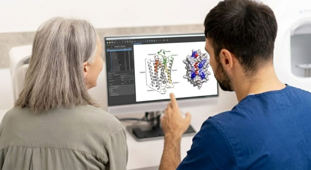
For you, this means that emerging molecular imaging technologies could help catch inflammatory or malignant changes earlier than traditional visual checks. While promising, these tools are currently designed to complement, not replace, standard assessments.
Keeping up with innovation puts you in control of your care. As these advanced diagnostics become more widely available, you may benefit from more personalised monitoring and treatment.
Speaking with a specialist ensures that any use of new technology is guided by evidence. Technology can enhance care, but your clinician’s judgement is still the key to safe and effective management.
FAQs:
1. What is molecular imaging in dermatology?
Molecular imaging is a technique that looks beneath the skin’s surface to detect cellular and biochemical activity. Unlike standard visual checks or structural scans, it reveals how tissue behaves at a molecular level, allowing clinicians to spot early inflammatory or malignant changes before they become visible.
2. How does it differ from traditional imaging?
Traditional imaging mainly shows structure size, thickness, or damage. Molecular imaging goes further by showing function, highlighting biochemical and cellular changes. This allows for earlier intervention and more precise treatment decisions.
3. Can molecular imaging detect skin cancer early?
Yes, it has the potential to detect abnormal cellular or metabolic activity before lesions appear visibly suspicious. This early insight may improve prognosis by enabling timely and targeted treatment.
4. How is molecular imaging useful for inflammatory skin conditions?
It can detect subclinical inflammation, cytokine activity, and microvascular changes before surface redness or plaques appear. For conditions like eczema and psoriasis, this may allow clinicians to adjust therapy earlier and more accurately.
5. Does molecular imaging replace biopsy?
No. Biopsy remains the gold standard for diagnosis. Molecular imaging can reduce unnecessary biopsies by highlighting suspicious areas and guiding sampling, making procedures more precise.
6. Can it guide biologic therapy decisions?
Potentially, yes. Imaging may show which inflammatory pathways are active before starting biologics, allowing more targeted treatment and better monitoring of response at a molecular level.
7. Is AI involved in molecular imaging?
Artificial intelligence analyses complex imaging data, recognising subtle patterns and supporting early detection. However, it complements rather than replaces clinical judgement. Your clinician remains central to interpreting results.
8. Are there limitations to current molecular imaging technology?
Yes. Most techniques are still in research or early clinical stages, with limited access and high cost. Accuracy, standardisation, and long-term clinical validation are ongoing, so it’s important to view the technology as complementary rather than definitive.
9. Is the data safe and private?
Advanced molecular imaging generates detailed biological information, so ethical data handling, informed consent, and robust privacy safeguards are essential. Transparency and regulatory compliance ensure your health information remains protected.
10. What does this mean for my care?
Molecular imaging may provide earlier detection of inflammatory or malignant changes, support personalised treatment plans, and help clinicians intervene proactively. While promising, it currently supplements standard assessments, with your specialist guiding all decisions.
Final Thoughts: Molecular Imaging and the Future of Dermatology
Molecular imaging represents a remarkable advance in dermatology, offering the potential to detect inflammatory and malignant changes beneath the skin before they become visible. By revealing cellular and biochemical activity, it moves care from reactive observation to proactive intervention. While the technology is still emerging and primarily complementary to traditional examinations and biopsies, its promise lies in enabling earlier detection, more precise treatment, and personalised management for conditions ranging from eczema and psoriasis to early-stage skin cancers.
If you’re thinking about a dermatologist in London, you can contact us at London Dermatology Centre to book a consultation with one of our specialists and explore how emerging molecular imaging technologies could support your skin health.
References:
- Berry, E., Thompson, R. F., Shachaf, C. & Leachman, S. (2025) Molecular Imaging in Early Skin Cancer Detection: Advances, Limitations, and Future Directions, Technology in Cancer Research & Treatment, https://pubmed.ncbi.nlm.nih.gov/41451881/
- Birmpili, A. (2025) From Optical to Molecular Imaging on Human Skin: A Review, Journal of Biomedical Imaging and Bioengineering, https://pmc.ncbi.nlm.nih.gov/articles/PMC12848729/
- Caruntu, C., Ilie, M.A. & Neagu, M. (2023) Looking into the Skin in Health and Disease: From Microscopy Imaging Techniques to Molecular Analysis, International Journal of Molecular Sciences https://www.mdpi.com/1422-0067/24/18/13737
- Gambichler, T., Jaedicke, V. & Stücker, M. (2019) Emerging imaging technologies in dermatology: Part I: Basic principles, Journal of the American Academy of Dermatology https://www.sciencedirect.com/science/article/abs/pii/S019096221832975X
- Vonk, J., de Wit, J.G., Witteman, C. et al. (2022) Fluorescence molecular imaging using cetuximab‑800CW in cutaneous squamous cell carcinoma surgery: a proof‑of‑concept study, British Journal of Dermatology, https://academic.oup.com/bjd/article/187/5/810/6966375