



If you have ever had a mole examined with a small illuminated magnifying device, you may already have experienced dermoscopy. What takes only moments during your skin examination developed through centuries of advances in microscopy, optics, illumination and understanding of structures beneath the skin’s surface.
Modern dermoscopy was not invented by one person at one particular moment. Your modern dermatoscope developed through contributions from many physicians and scientists, with especially important advances occurring during the nineteenth and twentieth centuries as the technique evolved from experimental microscopy into a practical method for assessing suspicious skin lesions.
What Is Dermoscopy?
Dermoscopy is a non-invasive technique that allows a trained clinician to examine the colours, structures and patterns within a skin lesion using a device called a dermatoscope. It provides a closer view of the skin and can help your clinician assess features that may not be obvious during an ordinary visual examination.
By combining magnification with specialised illumination, dermoscopy can reveal details beneath the skin’s surface more clearly. This additional information can support a more accurate assessment of moles and other skin lesions.
The Story Began With Early Microscopes
The earliest roots of dermoscopy can be traced to seventeenth-century experiments showing that magnification could reveal details of the skin and small blood vessels that were not visible to the naked eye. Pierre Borel published microscopic observations in 1655 and 1656, including early examination of human skin and nail-bed capillaries.
These experiments were not intended to diagnose skin cancer. Their historical importance lies in demonstrating that magnification could reveal anatomical details that ordinary visual examination could not show clearly.
Examining Tiny Blood Vessels
In 1663, Johan Christophorus Kohlhaus reproduced early microscopic observations of small vessels around the nail. His work therefore represents another early milestone in the long development of techniques used to examine structures beneath your skin’s visible surface.
You would not recognise these early microscopes as modern dermatoscopes. However, they demonstrated that magnification could reveal vascular details that could not be assessed reliably with your naked eye.
Digital Dermoscopy Changed Monitoring
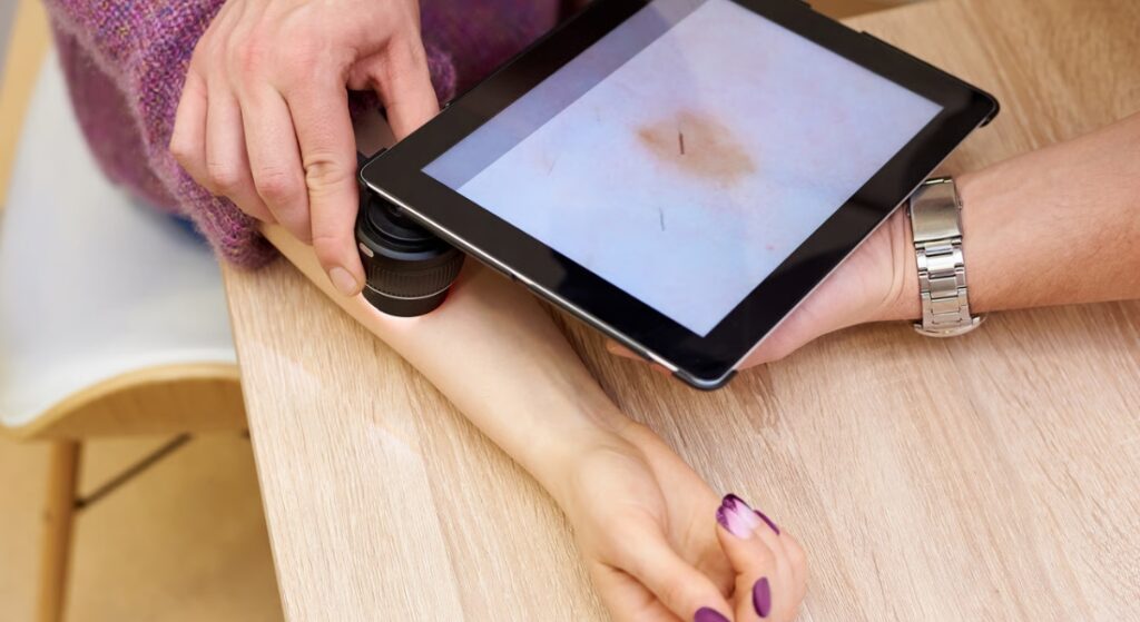
Digital dermoscopy made it possible for your dermatologist to record high-quality dermoscopic images and compare selected lesions at later appointments. This can help identify subtle structural changes that may be difficult to recognise from memory alone.
Digital monitoring is not necessary for every mole. In UK specialist care, NICE recommends baseline photography, preferably dermoscopic, when your clinically atypical melanocytic lesion does not initially require excision, followed by comparison after three months to look for early signs of melanoma.
Immersion Techniques Improved the View
During the nineteenth century, advances in immersion microscopy helped scientists understand how reducing reflection between optical surfaces could improve visualisation. Ernst Abbe’s work with immersion techniques in the 1870s formed part of this wider development in microscopy.
In 1893, dermatologist Paul Gerson Unna described diascopy and showed that manipulating the optical properties of the skin could make underlying colours and structures easier to see. These principles of reducing surface interference and improving visualisation helped lay foundations for later contact and immersion dermoscopy.
Portable Microscopes Made Examination Easier
Smaller monocular and binocular microscopes made examining skin under magnification more practical during clinical appointments. Doctors could study skin structures more directly while assessing patients.
- Smaller instruments: Microscopes became easier to use in clinical settings.
- Direct examination: Doctors could examine skin structures during patient appointments.
- Greater practicality: Smaller devices reduced reliance on large laboratory equipment.
- Clinical use: Magnified skin examination became more accessible in everyday practice.
These developments helped bring microscopic skin examination closer to routine clinical care. They also created a foundation for later advances in skin microscopy and dermoscopy.
Johann Saphier Gave the Technique a Name
Around 1920, Johann Saphier began publishing a series of papers in which he used the term dermatoskopie. He systematically examined structures beneath your skin using a binocular microscope and described vascular, pigmentary and other patterns.
Saphier’s work helped establish dermatoscopy as a recognisable clinical technique rather than simply an experimental use of microscopy. However, the method was not yet primarily being used to determine whether your pigmented lesion was melanoma.
Early Dermoscopy Was Not Mainly About Melanoma
Saphier used dermatoscopy to investigate normal and diseased skin, particularly vascular structures and pigmentation. Although he also examined melanocytic naevi, melanoma diagnosis was not yet the central purpose of your dermoscopic examination.
The shift towards skin-cancer assessment occurred gradually during later decades. This distinction is important because the modern association between dermoscopy and melanoma represents a later stage in the technique’s history.
Leon Goldman Advanced Pigmented-Lesion Research
During the 1950s, American dermatologist Leon Goldman applied skin-surface microscopy to your pigmented naevi and melanoma. His 1951 work helped move the technique towards the detailed assessment of melanocytic lesions, although the available equipment still had important limitations.
In 1958, Goldman also described a simple portable microscope for examining your skin. This represented another step towards making magnified skin examination more practical at the point of care.
The Dermatoscope Became More Practical
Goldman’s portable microscope was an important development, but the handheld dermatoscope you recognise today developed further during subsequent decades. Improvements in lenses, illumination and compact instrument design made detailed skin-surface examination increasingly practical.
A major milestone came in 1989 with the commercially available Heine Delta 10, based on work by Peter Bilek and Wilhelm Stolz. Your clinician could now use a compact illuminated device during a routine consultation rather than depending on cumbersome microscopy equipment.
Why Specialist Training Matters

Your dermatoscope provides additional visual information, but correctly interpreting the colours, structures and patterns requires training and clinical experience. Your dermatologist must consider these findings alongside your lesion’s appearance, location, history and any changes you have noticed.
If you see an experienced dermatologist in London for assessment of a suspicious lesion, dermoscopy may help determine whether the lesion appears reassuring, requires monitoring or needs tissue examination. When melanoma is suspected, complete excision biopsy is generally preferred where clinically appropriate so that the lesion can be assessed histologically, although the exact biopsy approach depends on the lesion and its location.
Melanoma Detection Became a Major Focus
During the 1970s, Rona MacKie helped show that subsurface features visible through skin microscopy could help distinguish melanocytic naevi from melanoma. This helped shift dermoscopy towards practical melanoma assessment, with further research in the following decade identifying colours, structures and patterns linked with benign and malignant lesions.
Development of Dermoscopy for Melanoma Detection
| Period | Development | Impact on Melanoma Assessment | Why It Mattered |
| 1970s | Rona MacKie studied subsurface features seen through skin microscopy | Helped distinguish melanocytic naevi from melanoma | Demonstrated the clinical potential of skin microscopy |
| Early research | Specialists examined colours and structures beneath the skin surface | Improved understanding of differences between lesions | Added greater detail to visual assessment |
| Pattern recognition | Researchers investigated combinations of colours, structures and patterns | Supported more systematic lesion assessment | Helped move beyond simple visual description |
| Benign lesions | Features associated with benign melanocytic naevi were studied | Improved recognition of lesions less likely to be malignant | Could help reduce unnecessary concern |
| Malignant lesions | Features associated with melanoma were investigated | Supported recognition of suspicious lesions | Strengthened the role of dermoscopy in melanoma assessment |
| Continued research | Evidence accumulated across multiple specialists and studies | Built the foundations of modern melanoma assessment | Dermoscopy developed through collective research rather than one discovery |
Pattern Analysis Changed How Lesions Were Assessed
In 1987, influential studies by Pehamberger, Steiner and Wolff described systematic pattern analysis for your pigmented skin lesions and investigated its value in identifying early melanoma. These approaches encouraged clinicians to assess combinations of dermoscopic structures rather than relying on one isolated feature.
Standardisation became increasingly important as the technique spread. A consensus meeting held in Hamburg in 1989 established terminology for skin-surface microscopy, with the recommendations published in 1990. Later algorithms and consensus exercises continued refining how your dermoscopic findings are described and interpreted.
Handheld Dermatoscopes Expanded Use
The development of compact illuminated dermatoscopes made the technique much easier to incorporate into your routine dermatology consultation. The Heine Delta 10 became commercially available in 1989 and helped establish the basic format of the portable dermatoscope used widely thereafter.
Modern devices commonly provide approximately tenfold magnification with integrated illumination. Your dermatologist can therefore examine a lesion immediately rather than requiring a separate large microscopy system.
Polarised Light Brought Another Advance
Later dermatoscopes introduced cross-polarised illumination, which reduces surface glare without always requiring direct contact or immersion fluid. Your dermatologist may therefore be able to examine deeper dermoscopic structures conveniently while avoiding pressure on the lesion.
Polarised and non-polarised dermoscopy do not produce identical images. Some structures are more conspicuous with one technique than the other, so your dermatologist may use a device capable of switching between them when this provides useful additional information.
Why Dermoscopy Is Important Today

Dermoscopy is now an established part of specialist melanoma assessment. NICE recommends that your suspected melanoma should be examined with dermoscopy during specialist assessment by a healthcare professional trained in the technique.
When used by trained specialists, dermoscopy can improve melanoma recognition compared with naked-eye inspection alone and may reduce unnecessary removal of benign lesions. Its role has also expanded to basal cell carcinoma and many inflammatory, infectious, hair, nail and other dermatological conditions.
Myth vs Fact
| Myth | What You Should Know |
| One person invented the dermatoscope you use today. | Your modern dermoscopy developed through contributions from many physicians and optical researchers over several centuries. |
| The first skin microscopy was developed specifically to detect melanoma. | Your earliest microscopy focused on structures such as blood vessels rather than skin cancer. |
| Johann Saphier invented the complete modern technique. | Saphier gave dermatoscopy its name and made major contributions, but your modern technique developed through many later advances. |
| Dermoscopy became a melanoma tool immediately. | Your melanoma-focused dermoscopy developed particularly during the second half of the twentieth century. |
| A dermatoscope can tell your dermatologist with certainty that you have cancer. | Your dermoscopy improves assessment but histological examination may still be needed for diagnosis. |
| Digital dermoscopy means every mole should simply be monitored. | Your suspicious lesion may require biopsy or excision rather than monitoring. |
| Polarised and non-polarised dermoscopy show exactly the same structures. | Your dermatologist may see somewhat different features with the two optical techniques. |
| Anyone using a dermatoscope gets the same diagnostic benefit. | Your interpretation depends substantially on appropriate training and clinical expertise. |
Key Takeaways
- Your modern dermatoscope developed gradually rather than being invented by one person.
- The earliest roots of your dermoscopic examination can be traced to microscopy experiments during the seventeenth century.
- Pierre Borel described early microscopic observations in 1655–1656, followed by Johan Christophorus Kohlhaus in 1663.
- Paul Gerson Unna helped establish the importance of reducing surface reflection when examining your skin.
- Johann Saphier introduced the term dermatoskopie around 1920.
- Leon Goldman helped extend skin-surface microscopy to your pigmented lesions during the 1950s.
- Rona MacKie helped establish dermoscopy’s potential for distinguishing melanoma from benign melanocytic lesions during the 1970s.
- Pattern analysis and standardised terminology made your dermoscopic assessment more systematic during the 1980s and 1990s.
- Portable handheld dermatoscopes helped bring the technique into routine clinical practice.
- Your dermoscopy can improve specialist assessment of suspicious lesions but cannot replace histopathology when tissue examination is required.
UK Guidance Note
If your pigmented lesion is referred for assessment or identified during melanoma follow-up in secondary or tertiary care, NICE recommends that it should be examined using dermoscopy by a healthcare professional trained in the technique. Your dermoscopic assessment should form part of a wider clinical evaluation rather than being treated as an independent cancer test.
If a lesion remains suspicious, your specialist may recommend excision biopsy or another appropriate method of obtaining tissue for histological examination. You should seek professional assessment if you notice a new, changing or unusual lesion rather than trying to determine whether it is benign or malignant from photographs alone.
Frequently Asked Questions
1. Who invented the dermoscopy used for your skin examination?
No single person invented your modern dermoscopy. Its development involved centuries of advances, including work by Pierre Borel, Paul Gerson Unna, Johann Saphier, Leon Goldman, Rona MacKie and many later researchers.
2. When did the history of your dermoscopy begin?
The earliest optical roots can be traced to seventeenth-century microscopy, including Pierre Borel’s work in 1655–1656 and observations reproduced by Johan Christophorus Kohlhaus in 1663. Modern clinical dermoscopy developed much later.
3. Who introduced the word dermatoscopy?
Johann Saphier introduced dermatoskopie in papers beginning around 1920. His work helped establish your skin-surface microscopy as a distinct clinical technique.
4. When did dermoscopy begin to help detect melanoma?
Work during the second half of the twentieth century gradually established this role. Rona MacKie’s research during the 1970s was particularly important in demonstrating how subsurface patterns could help distinguish your benign melanocytic lesion from melanoma.
5. When did handheld dermatoscopes become available?
Portable instruments developed progressively during the twentieth century, with an important milestone occurring in 1989 when the Heine Delta 10 became commercially available. Your modern handheld devices developed further from this type of design.
6. What does dermoscopy show that a normal examination may not?
Your dermatoscope combines magnification and specialised illumination to reveal colours and structures in the epidermis and superficial dermis that may not be apparent to your naked eye.
7. What is digital dermoscopy?
Your digital dermoscopy allows detailed lesion images to be stored and compared over time. Your dermatologist may use serial images for selected lesions when monitoring is clinically more appropriate than immediate removal.
8. Can dermoscopy diagnose your skin cancer with certainty?
No. Your dermoscopy can substantially improve assessment of a suspicious lesion, but it does not replace histological examination when biopsy or excision is needed to establish a tissue diagnosis.
9. Can dermoscopy help with conditions other than melanoma?
Yes. Your dermatologist may use dermoscopy when assessing basal cell carcinoma and other skin cancers as well as inflammatory, infectious, hair, nail and other non-cancerous conditions.
10. Why does your dermatologist need training in dermoscopy?
Your dermatoscope reveals patterns that require appropriate interpretation. Your dermatologist needs to combine these findings with your clinical history, the lesion’s appearance and other examination findings when deciding whether monitoring, biopsy or treatment is appropriate.
Final Thoughts: The Evolution of Dermoscopy
Your modern dermoscopic examination developed through centuries of advances in microscopy, optics and dermatology rather than through one invention. From early observations of tiny blood vessels to Saphier’s dermatoskopie, melanoma pattern analysis, handheld instruments and digital imaging, each development helped make examination of structures beneath your skin more practical and clinically useful. If you’re looking for an experienced dermatologist in London for assessment of a mole or suspicious skin lesion, you can get in touch with us at London Dermatology Centre.
References:
- Brancaccio, G., Russo, T., Lallas, A., Moscarella, E., Agozzino, M. and Argenziano, G. (2017) ‘Melanoma: clinical and dermoscopic diagnosis’, Giornale Italiano di Dermatologia e Venereologia, 152(3), pp. 213–223. Available at: https://pubmed.ncbi.nlm.nih.gov/28121084/
- Buch, J. and Criton, S. (2021) ‘Dermoscopy Saga – A Tale of 5 Centuries’, Indian Journal of Dermatology, 66(2), pp. 174–178. Available at: https://pmc.ncbi.nlm.nih.gov/articles/PMC8208256/
- Rao, B.K. and Ahn, C.S. (2012) ‘Dermatoscopy for melanoma and pigmented lesions’, Dermatologic Clinics, 30(3), pp. 413–434. Available at: https://www.sciencedirect.com/science/article/pii/S073386351200037X
- Rajpara, S.M., Botello, A.P., Townend, J. and Ormerod, A.D. (2009) ‘Systematic review of dermoscopy and digital dermoscopy/artificial intelligence for the diagnosis of melanoma’, British Journal of Dermatology, 161(3), pp. 591–604. Available at: https://pubmed.ncbi.nlm.nih.gov/19302072/
- Marghoob, A.A. (2026) ‘A Window Beneath the Skin: The History of Dermoscopy from Curiosity to Clinical Cornerstone’, Dermatology Practical & Conceptual, 16(1), article 7121. Available at: https://pmc.ncbi.nlm.nih.gov/articles/PMC12952884/