")
You’ll often assume that any flat brown mark on sun-exposed skin is a liver spot, simply because they’re so common and familiar. They do tend to appear in predictable places over time, but appearances can be deceptive. Not every dark patch is the same, and even subtle differences can point to entirely different causes.
Several other skin conditions can mimic liver spots, some harmless and others needing attention. That’s why relying purely on visual cues can mislead you, especially if you’re trying to manage them at home. Without professional input, it’s easy to either overreact or overlook something that should be monitored more closely.
Knowing the distinctions matters for both peace of mind and safety. Understanding what could be mistaken for liver spots helps you avoid unnecessary worry while making informed decisions about treatment or assessment. When you combine awareness with professional guidance, you ensure nothing important is missed and that your approach remains targeted and effective.
What Are Liver Spots Supposed to Look Like?
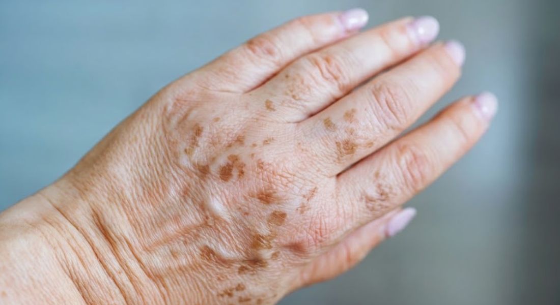
You’ll notice that liver spots, or solar lentigines, are generally flat and evenly coloured, making them relatively easy to identify once you know what to look for. Their tones range from light brown to dark brown, and they usually have clearly defined edges. They most often appear on areas that get frequent sun exposure, such as your face, hands, shoulders, and arms.
They develop slowly over time rather than appearing suddenly, and you may see an increase in number as cumulative sun exposure builds. Individual spots typically remain stable in both shape and size, so while the overall pattern changes gradually, each mark is fairly predictable in its appearance.
One of the key points is that liver spots aren’t raised or textured they remain smooth to the touch and don’t cause discomfort. They don’t itch, bleed, or change rapidly, which is what sets them apart from other skin conditions that might look similar at first glance.
Freckles vs Liver Spots
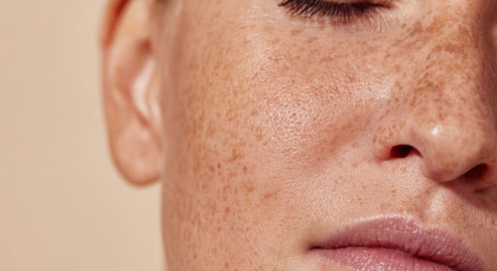
You’ll quickly notice that freckles and liver spots can look similar at first, but they behave very differently. Freckles are typically smaller, more numerous, and tend to appear earlier in life, often during childhood or adolescence. That makes them more transient in nature compared with the deeper, more persistent liver spots.
The biggest difference is how they react to the environment. Freckles often fade during periods of reduced sun exposure, while liver spots hold their colour regardless of the season. That persistence is a defining feature and is usually what tips you off that you’re dealing with liver spots rather than freckles.
Genetics also plays a bigger role with freckles, shaping where and how many appear. While sun exposure still influences both, freckles respond more noticeably to changes in your environment, which helps you distinguish them over time. Liver spots, by contrast, reflect cumulative exposure and remain stable once they’ve formed.
Melasma and Hormonal Pigmentation
You’ll see that melasma can look similar to liver spots, but the presentation is noticeably different. Rather than individual, well-defined spots, melasma appears as larger patches with softer, less distinct borders. That difference in shape and spread is usually the first clue that you’re dealing with hormonal pigmentation rather than sun-induced spots.
Hormones play a central role in melasma, which is why it often shows up during pregnancy or with certain hormonal treatments. Liver spots, by contrast, are primarily linked to cumulative sun exposure and aren’t driven by hormonal shifts. That distinction is key when deciding how to manage or treat the pigmentation effectively.
The pattern and location also help differentiate melasma from liver spots. It tends to affect the cheeks, forehead, and upper lip in a symmetrical way, whereas liver spots are more isolated and random across sun-exposed areas. Understanding these differences ensures you can target the right condition with the most appropriate approach.
Post-Inflammatory Hyperpigmentation
Post-inflammatory hyperpigmentation can be confusing because it often looks a lot like liver spots. You might notice flat brown marks after a breakout, a cut, or any irritation, and wonder why they appeared in the first place. The difference is that these spots have a story you can usually link them to a recent skin event, which makes them easier to manage.
- Track the Cause: Take a moment to identify what triggered the mark in the first place. Whether it was acne, a scratch, or friction from clothing, knowing the origin helps you avoid repeating the same irritation and gives context for how the skin might heal.
- Gentle Skincare Is Essentia: Resist the urge to scrub aggressively or use harsh acids to “speed up” fading. These actions can actually worsen pigmentation and prolong healing. Stick to soothing, non-comedogenic products that calm the skin while supporting its natural repair process.
- Targeted Lightening Treatments Help: Ingredients like vitamin C, niacinamide, or azelaic acid can gradually reduce pigmentation when applied consistently. Overloading the skin with multiple actives at once can backfire, so it’s better to focus on one or two key treatments and monitor progress carefully.
- Sun Protection Prevents Darkening: Even brief exposure to sunlight can deepen post-inflammatory marks, making them more stubborn. Daily SPF is non-negotiable, and reapplying when you’re outdoors ensures your skin has the best chance to fade evenly without extra pigmentation forming.
- Patience Pays Off: Unlike liver spots, these marks often fade naturally over time. It’s tempting to jump from one product to another, but giving your skin several weeks to respond is the only way to truly see which approach works and avoid unnecessary irritation.
When you take the right stepsidentifying triggers, protecting your skin, and applying targeted treatments consistently you can significantly reduce these marks. Acting early and staying patient gives you the best chance of fading them completely, leaving your skin looking even and healthy.
Seborrhoeic Keratoses
Seborrhoeic keratoses can catch you off guard because they often look very similar to liver spots at first glance. You might notice them starting as small, flat patches that gradually become raised, with a waxy or rough surface that feels different from the smoothness of typical age spots. The colour can range from light brown to almost black, and up close, the texture is usually the giveaway that these aren’t your usual sun spots.
You shouldn’t panic if you spot one, because they’re entirely benign. What’s tricky is that they can mimic more serious skin changes, so relying solely on appearance can be misleading. You might feel tempted to ignore them, but getting a proper assessment ensures that nothing dangerous is missed.
While they don’t require treatment for health reasons, you may choose to have them removed if they catch on clothing or simply bother you cosmetically. The key is knowing the difference between harmless growths and changes that need closer inspection you don’t want to miss something important while worrying over harmless bumps.
Actinic Keratosis and Sun Damage
Actinic keratosis can easily be mistaken for a simple liver spot at first, especially when it starts as a small, flat patch on sun-exposed areas like your face, hands, or forearms. You might not notice anything unusual initially, but over time, the surface often becomes rough or scaly, setting it apart from the smooth appearance of typical sun spots. This texture is usually the first clue that something needs closer attention.
It’s important to treat these lesions seriously because they’re considered precancerous. You don’t have to panic, as not every case will progress, but regular monitoring is essential to catch any changes early. You should be proactive about having suspicious patches evaluated rather than waiting for them to worsen.
If you’re unsure, touch can be a helpful guide. Actinic keratosis often feels different to the touch rough, dry, or flaky compared with the surrounding skin. Any lesion that changes in size, texture, or colour should be checked professionally; early intervention makes a big difference in preventing complications.
Lentigo Maligna and Skin Cancer Risk
Lentigo maligna can be deceptive because it often starts as a flat, pigmented patch that looks almost identical to a harmless liver spot. You might not notice anything alarming at first, especially if it sits quietly on sun-exposed skin like your face or hands. The danger is that its early appearance masks a serious risk, which is why awareness is key.
Over time, you may see subtle changes in shape, colour, or texture. The edges can become uneven or blurred, and the pigmentation may darken or lighten in different areas. These shifts are critical warning signs that shouldn’t be overlooked you want to catch them before they develop into a more invasive melanoma.
Because lentigo maligna mimics benign spots so closely, you can’t rely on sight alone. Professional assessment is crucial, and early detection dramatically improves the outcome. If you notice any new spot behaving differently, don’t wait get it checked promptly to reduce the risk of progression.
Moles and Pigmented Lesions
Moles can easily throw you off because some are flat and pigmented, making them look a lot like liver spots at first glance. You might assume they’re harmless, especially if they’ve been there for years, but the key is to pay attention to subtle differences in colour and shape. Their uniformity and well-defined edges are usually reassuring, but changes can happen gradually.
You need to keep an eye on them over time. Any shift in size, colour, or border can signal that a mole warrants closer inspection. Regular self-checks help you distinguish between the harmless ones and those that could pose a risk.
Unlike liver spots, moles aren’t purely a result of sun exposure; genetics often play a significant part. This distinction matters when assessing risk and planning any necessary intervention. Knowing the source helps you and your dermatologist decide whether monitoring, removal, or further investigation is needed.
Why Visual Similarity Can Be Misleading
It’s easy to assume a brown spot is just a liver spot, but appearances can be deceiving. You might notice a mark on your skin and think it’s harmless, yet the colour, shape, or location could point to a completely different issue. Even trained eyes can struggle to differentiate between similar-looking conditions without considering other factors.
- Lighting and Skin Tone Affect Perception: The way a spot looks can change dramatically depending on the light and your natural skin tone. A mark that seems flat or faint in one setting may appear darker or more pronounced in another, which can make self-assessment unreliable.
- Location Influences Interpretation: Spots on sun-exposed areas behave differently than those in shaded regions. You might assume a cheek mark is a liver spot, but it could be post-inflammatory pigmentation from acne or irritation, especially if it follows a pattern.
- Similar Colours Don’t Equal Similar Causes: Brown, beige, or reddish spots often share visual traits, but the underlying cause varies widely. Treating a spot based solely on how it looks can delay proper care or even make it worse if the wrong approach is used.
- Professional Assessment Considers Context: A dermatologist looks at more than colour and shape; they check history, texture, and associated symptoms. This holistic approach reduces errors and ensures you get treatment that actually works.
- Assumptions Lead to Mistakes: Jumping to conclusions about a spot’s identity is risky. Misdiagnosis can result in unnecessary treatments or overlooked conditions, which is why taking a cautious, informed approach matters most.
When you remember that spots are more than just their appearance, you can avoid costly mistakes and take steps that actually benefit your skin. Checking the history, pattern, and context of each mark gives you clarity and confidence in managing them.
The Importance of Skin History
Your skin history is more important than you might think when figuring out what a spot really is. You should consider when it first appeared and whether it has changed, because timing often gives more insight than appearance alone. A sudden spot or one that has shifted over months can point to something other than a typical liver spot.
You also need to factor in what might have triggered the mark. Spots linked to past irritation, injury, or inflammation usually have a different cause than sun-related pigmentation. Understanding the context of each mark helps you separate harmless changes from those that need attention.
Keeping a record of your spots makes monitoring far easier. Simple photographs over weeks or months can reveal subtle changes that might be missed otherwise. This evidence is invaluable when you consult a professional, making assessment quicker and more accurate.
When Changes Should Raise Concern

You should pay attention if a spot changes noticeably, because certain shifts can signal something more serious. Rapid growth, uneven edges, or multiple colours within a single area are all red flags. You should also be alert to symptoms like bleeding, itching, or tenderness these aren’t typical for standard liver spots.
Liver spots usually stay the same over time, so any variation is worth noting. You shouldn’t assume that stability is guaranteed; even small deviations can be important. Getting an early professional assessment ensures you catch anything concerning before it progresses.
Ultimately, trusting your instincts matters. If a spot feels off or just doesn’t look right, don’t wait it out. Acting early is the safest approach and gives you the best chance of addressing potential problems effectively.
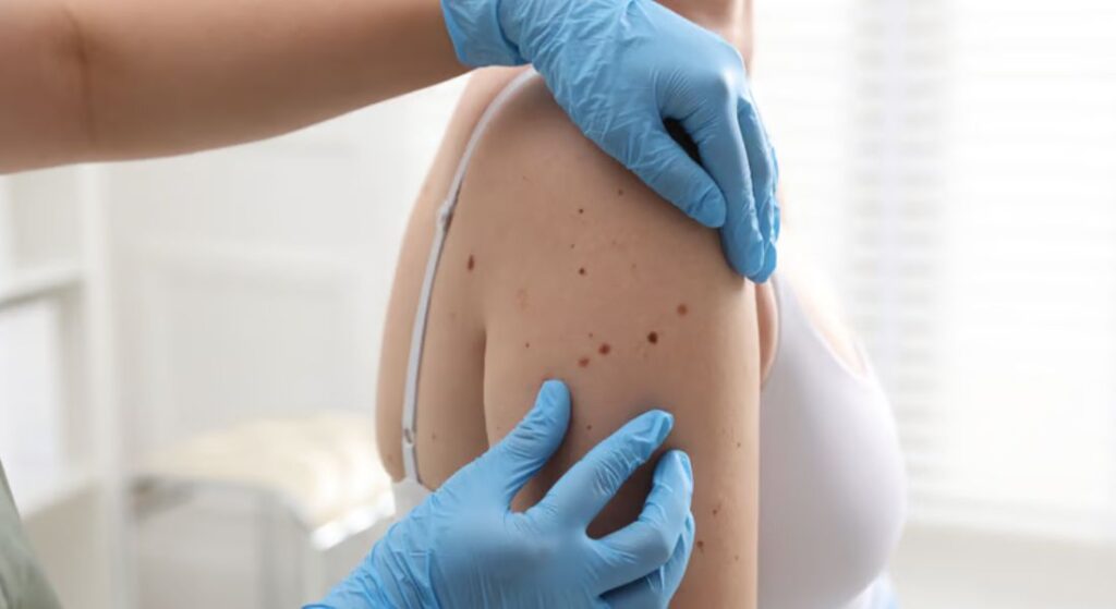
How Dermatologists Differentiate Skin Lesions
Dermatologists have the advantage of tools and expertise that make spotting the difference between lesions far easier than you can do at home. Dermo copy, for example, lets them see pigment patterns and structures beneath the surface that are invisible to the naked eye. This level of detail helps separate harmless liver spots from potentially serious lesions.
Sometimes, visual inspection alone isn’t enough. In those cases, a biopsy can confirm exactly what a spot is. You might worry about the procedure, but it’s quick, precise, and provides definitive answers that guide the next steps.
Relying on professional assessment removes all the guesswork. You gain clarity and reassurance, and any necessary treatment can be planned accurately. For you, that means peace of mind and a clear path forward without leaving anything to chance.
Why Self-Diagnosis Can Be Risky
Self-diagnosis is common but not always reliable. Many online resources provide general descriptions that may not apply to your specific case. This can lead to confusion or false reassurance.
Some serious conditions can initially look harmless. Without proper training, it is easy to miss subtle warning signs. This is why relying solely on self-assessment can be risky.
Seeking professional advice ensures that nothing is overlooked. It allows for early detection and appropriate management. This is particularly important for pigmented lesions.
Preventing Misdiagnosis Through Awareness
Being aware that not every brown spot is a liver spot is a powerful first step in avoiding misdiagnosis. You should approach new or changing marks with caution rather than assuming they’re harmless. This mindset helps you notice changes that might otherwise go unchecked.
Familiarising yourself with the typical signs of different lesions can guide your observations, but it’s not a substitute for professional evaluation. You can use knowledge to make smarter decisions about when to seek help, spotting red flags earlier and reducing unnecessary worry.
Proactivity pays off when it comes to skin health. Regular self-checks, combined with timely dermatology consultations, improve outcomes and allow early intervention where needed. Staying vigilant ensures you catch issues before they escalate, keeping your skin safer over the long term.
FAQs:
1. What exactly are liver spots?
Liver spots, also called solar lentigines, are flat, brown patches that appear on sun-exposed skin. They are usually harmless and develop slowly over time, remaining stable in size and shape.
2. Can freckles be mistaken for liver spots?
Yes, freckles can look similar, but they are generally smaller, more numerous, and fade when sun exposure decreases. Liver spots persist regardless of the season.
3. How is melasma different from liver spots?
Melasma appears as larger, irregular patches with soft, blurred edges, often linked to hormonal changes. Liver spots are smaller, well-defined, and primarily caused by cumulative sun exposure.
4. What is post-inflammatory hyperpigmentation?
These are brown marks that appear after acne, cuts, or skin irritation. Unlike liver spots, they often fade over time and are linked to a specific skin event.
5. Are seborrhoeic keratoses dangerous?
No, seborrhoeic keratoses are benign growths. They may resemble liver spots but often have a waxy, rough texture and can become slightly raised.
6. Should I worry about actinic keratosis?
Actinic keratosis is considered precancerous. While it can look like a simple liver spot at first, its rough, scaly texture and sun-exposed location mean it should be monitored and treated if necessary.
7. How can I tell a mole apart from a liver spot?
Moles are often pigmented and can be flat or raised. Changes in size, colour, or borders over time signal the need for closer inspection, unlike stable liver spots.
8. Is self-diagnosis of liver spots reliable?
Not always. Similar-looking spots can have different causes, and self-assessment may overlook subtle warning signs. Professional evaluation is safer.
9. When should I see a dermatologist?
Seek advice if a spot changes in size, shape, colour, texture, or starts bleeding or itching. Early assessment reduces risk and ensures correct treatment.
10. How can I prevent misdiagnosis?
Track your skin history, observe changes, protect your skin from sun exposure, and consult a professional when unsure. Awareness combined with expert guidance is key.
Final Thought: Staying Informed for Healthier Skin
Understanding that not every brown spot is a liver spot is essential for keeping your skin healthy and avoiding unnecessary worry. By learning to distinguish between freckles, melasma, post-inflammatory marks, seborrhoeic keratoses, actinic keratosis, and other pigmented lesions, you can make more informed decisions about your skin care and know when professional assessment is necessary. Regular monitoring, sun protection, and awareness of changes in your skin are your first lines of defense. If you’re looking for liver spots treatment in London, you can reach out to us at the London Dermatology Centre to book a consultation with one of our specialists. This proactive approach ensures that any concerns are addressed early, giving you peace of mind and a clear plan for maintaining healthy, even-toned skin.
References:
- Elgart, G.W. (2001) Seborrheic keratoses, solar lentigines, and lichenoid keratoses: dermatoscopic features and correlation to histology and clinical signs. Dermatologic Clinics, 19(2), pp.347–357. Available at: https://pubmed.ncbi.nlm.nih.gov/11556243/
- Mardani, G., Nasiri, M.J., Namazi, N., Farshchian, M. & Abdollahimajd, F. (2025) Treatment of Solar Lentigines: A Systematic Review of Clinical Trials. Journal of Cosmetic Dermatology, 24(4), e70133. Available at: https://pubmed.ncbi.nlm.nih.gov/40145274/
- Fitzpatrick, R.E., Goldman, M.P., Ruiz‑Esparza, J. & Marcos, J.D. (2015) Evaluation of pulsed dye laser (PDL) and other physical modalities for treatment of solar lentigines. Journal of Clinical and Aesthetic Dermatology (PMCID: PMC4281975). Available at: https://pmc.ncbi.nlm.nih.gov/articles/PMC4281975/
- Ortonne, J.P., Pandya, A.G. & Hexsel, D. (2006) Treatment of solar lentigines: a review of physical and topical therapies, Journal of the American Academy of Dermatology, 54(5 Suppl 2), S262–S271. Available at: https://www.sciencedirect.com/science/article/abs/pii/S0190962205049698
- Almond‑Roesler, B. & Zouboulis, C.C. (2000) Successful treatment of solar lentigines by brief gentle cryosurgery using a Kryomed device, British Journal of Dermatology, 143(1), pp.216–218. Available at: https://academic.oup.com/bjd/article-abstract/143/1/216/6688206