


Basal cell carcinoma (BCC) is the most common type of skin cancer, affecting millions of people worldwide every year. Although it rarely spreads to distant parts of the body, it can continue to grow and damage nearby skin, cartilage, nerves and other important structures if left untreated. If you notice a persistent skin lesion or an unusual growth, seeking early assessment can help you receive prompt and effective treatment.
Most basal cell carcinomas can be successfully treated with surgery, but not every tumour behaves in the same way. A facial location can increase treatment complexity, particularly in areas such as the eyelids, nose, ears or lips. However, risk also depends on tumour size, borders, histological subtype, previous treatment, immune status and other clinical factors. These features increase the likelihood of the cancer returning if it is not completely removed.
Research comparing Mohs micrographic surgery with standard surgical excision has investigated which approach provides the best balance between tumour clearance, tissue preservation, cosmetic outcomes and long-term disease control. The study aims to determine which surgical approach provides the best balance between complete tumour removal, preservation of healthy tissue, cosmetic results and long-term disease control. As research continues, the findings may help your specialist make more informed treatment decisions and improve the care you receive.
What Is the OCTOPUS Trial?
The OCTOPUS trial is a clinical research study evaluating surgical treatment options for high-risk basal cell carcinoma (BCC). If you are diagnosed with a high-risk BCC, the study aims to improve understanding of which surgical approach offers the most effective treatment while reducing the risk of the cancer returning.
Researchers are comparing Mohs micrographic surgery with conventional surgical excision to assess how well each technique removes the tumour. They are also examining whether one approach provides better preservation of healthy tissue and improved cosmetic outcomes for you after surgery.
The aim of these comparative studies is to understand which surgical approach provides the best long-term outcomes for selected patients with high-risk basal cell carcinoma.The findings are expected to help your specialist choose the most appropriate surgical option for your individual tumour while supporting future clinical guidelines for the management of high-risk basal cell carcinoma.
Why Was the Study Needed?
Although both Mohs micrographic surgery and standard surgical excision are widely used to treat basal cell carcinoma, questions remain about which approach provides the greatest benefit for high-risk tumours. If you have a high-risk BCC, choosing the most appropriate surgical treatment can be important for reducing the chance of the cancer returning while achieving the best possible cosmetic result.
Researchers wanted stronger clinical evidence to compare these two surgical techniques under carefully controlled conditions. They aimed to understand which approach offers you the best balance between complete tumour removal, preservation of healthy tissue and long-term disease control.
The OCTOPUS trial was developed to address this important question in skin cancer management. Its findings are expected to help your specialist make more informed treatment decisions and provide evidence-based care that is tailored to your individual needs.
What Makes a Basal Cell Carcinoma High Risk?
Not all basal cell carcinomas behave in the same way, and certain features increase the likelihood of the cancer returning after treatment. If your tumour has characteristics associated with a higher risk of recurrence, your specialist may recommend a more specialised treatment approach to achieve the best possible outcome.
These high-risk features include a large tumour size, a location on the face or other sensitive areas, recurrent disease, aggressive microscopic subtypes and poorly defined tumour margins. Each of these factors can make it more difficult to remove the cancer completely while preserving healthy tissue.
High-risk basal cell carcinomas often require careful treatment planning and close follow-up. Your specialist will assess the features of your tumour before recommending the surgical option that is most appropriate for you, helping to reduce the risk of recurrence and achieve the best functional and cosmetic result.
Understanding Basal Cell Carcinoma
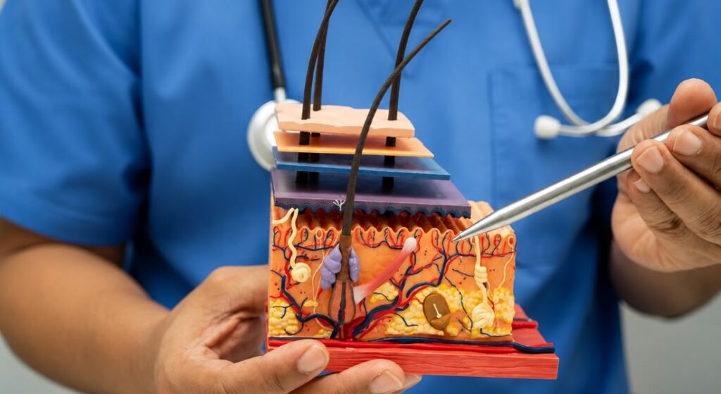
Basal cell carcinoma (BCC) develops from cells in the deepest layer of your epidermis, which is the outer layer of your skin. If you develop this type of skin cancer, it usually begins as a small lesion that changes gradually over time rather than causing sudden symptoms.
Most basal cell carcinomas grow slowly, but they can continue to spread into nearby skin and surrounding tissues if treatment is delayed. Although BCC rarely spreads to distant parts of your body, it can become locally destructive and may damage important structures if left untreated.
Fortunately, early diagnosis often leads to excellent outcomes. If you have your skin checked promptly and receive appropriate treatment, your specialist can usually remove the cancer successfully while helping to preserve as much healthy tissue as possible.
Factors That Influence the Choice Between Mohs Surgery and Standard Excision
| Tumour or patient factor | Why it matters | When Mohs surgery may be considered | When standard excision may remain appropriate |
| Tumour location | Surgery near the eyelids, nose, lips and ears may affect important cosmetic or functional structures. | Mohs may be useful where complete margin assessment and tissue preservation are especially important. | Excision may be suitable when clear margins can be achieved without unacceptable functional or cosmetic impact. |
| Primary or recurrent tumour | A recurrent BCC may have irregular extensions beyond the visible lesion and may be harder to remove completely. | Mohs is often considered for recurrent facial BCC because of its margin-control method and lower recurrence rates in randomised long-term evidence. | Standard excision may be considered in selected recurrent cases following specialist assessment. |
| Clinical borders | Poorly defined borders make it difficult to estimate the true extent of the cancer. | Staged microscopic examination can help identify tumour extending beyond the visible edge. | Excision is more straightforward when the tumour is well defined and an appropriate margin can be taken. |
| Histological subtype | Infiltrative, morphoeic, micronodular and other aggressive growth patterns may extend unpredictably into surrounding tissue. | Mohs may be considered when the subtype is associated with a greater risk of incomplete removal or recurrence. | Excision may remain suitable for well-defined, lower-risk histological subtypes. |
| Tumour size | Larger tumours may require wider removal and more complex reconstruction. | Mohs may help limit unnecessary tissue removal in anatomically constrained sites. | Standard excision may be appropriate when a suitable margin and repair can be achieved safely. |
| Previous incomplete excision | Cancer cells at or close to the surgical margin indicate that residual tumour may remain. | Mohs can map and remove remaining tumour while examining the margins during the procedure. | Re-excision may be reasonable when the area and likely residual tumour can be managed with an appropriate margin. |
| Perineural involvement | Tumour growth around nerves may increase the risk of recurrence and require specialist multidisciplinary management. | Mohs or another form of comprehensive margin-controlled surgery may form part of treatment in selected cases. | Standard excision alone may be insufficient when clinically significant nerve involvement is suspected or confirmed. |
| Immunosuppression | Immune suppression can affect tumour behaviour, healing and the risk of developing further skin cancers. | A margin-controlled approach may be considered when other high-risk features are also present. | Treatment still depends on tumour characteristics, general health and multidisciplinary advice. |
| Ability to tolerate a longer procedure | Mohs may involve several stages and a visit lasting several hours. | It may be appropriate when the patient can tolerate staged surgery under local anaesthetic. | Standard excision may be more practical where a shorter planned procedure is clinically suitable. |
| Local expertise and resources | Mohs requires trained surgeons, on-site tissue processing and close coordination between surgery and histology. | It should be performed in an appropriately equipped specialist service. | Standard excision is more widely available and remains effective for many appropriately selected BCCs. |
What Is Standard Surgical Excision?
Standard surgical excision is a common treatment for basal cell carcinoma that involves removing the visible tumour together with a margin of surrounding healthy tissue. If you undergo this procedure, your specialist aims to remove the entire cancer while reducing the risk of any tumour cells being left behind.
After the operation, the removed tissue is sent to a laboratory where the margins are examined under a microscope. This assessment helps determine whether the cancer has been completely removed or if tumour cells are still present at the edges of the specimen.
If cancer cells are found at the margins, you may require additional treatment or another surgical procedure to remove the remaining tumour. Your specialist will discuss the results with you and recommend the most appropriate next steps to ensure the best possible long-term outcome.
What Is Mohs Micrographic Surgery?
Mohs micrographic surgery is a specialised technique used to remove certain skin cancers with a high level of precision. If you have a high-risk basal cell carcinoma, your specialist may recommend this procedure because it aims to remove all of the cancer while preserving as much healthy skin as possible.
During the procedure, the tumour is removed in carefully controlled stages, with each thin layer of tissue examined immediately under a microscope. This allows your specialist to check whether cancer cells remain before deciding if any additional tissue needs to be removed.
The process continues until no cancer cells are detected in the examined tissue. By checking the margins during the operation, Mohs surgery may help preserve healthy tissue in selected cases, which can support reconstructive planning. However, the final scar and appearance depend on the extent of the tumour, the number of stages required, the repair method and individual healing.
Why Mohs Surgery Is Different
Mohs micrographic surgery is different from standard surgical excision because it allows the surgical margins to be checked during the procedure itself. This means your specialist can identify whether cancer cells remain before completing treatment, rather than waiting for results after surgery.
- Immediate Margin Checking: Tissue samples are examined during surgery to confirm whether cancer cells are still present.
- Precise Tumour Removal: Mohs surgery removes cancerous tissue while preserving as much healthy skin as possible.
- Tissue Preservation Benefits: Removing only affected areas can help reduce unnecessary damage to surrounding normal tissue.
- Useful in Sensitive Areas: It is particularly valuable for areas such as the nose, eyelids, lips, and ears where appearance and function are important.
Overall, Mohs surgery offers a highly precise approach to treating certain skin cancers while preserving healthy tissue. This makes it especially useful in delicate or visible areas where maintaining appearance and function is a priority. Your specialist can advise whether Mohs surgery is the most appropriate option for your specific skin cancer.
Complete Tumour Removal
One of the primary aims of the OCTOPUS trial is to compare how effectively Mohs micrographic surgery and standard surgical excision achieve complete tumour removal. If your basal cell carcinoma is removed completely during the first procedure, you are less likely to require additional treatment in the future.
Achieving complete tumour clearance is important because it reduces the risk of the cancer returning after surgery. Removing all cancer cells while preserving as much healthy tissue as possible is a key goal, particularly for high-risk basal cell carcinomas.
Successful surgery provides the best opportunity for long-term disease control. Findings from clinical research comparing these surgical approaches may help your specialist make more informed treatment decisions.
Preserving Healthy Tissue
Preserving healthy skin is especially important when treating basal cell carcinomas on the face and other visible areas. If your tumour is close to important structures such as your eyes, nose or lips, your specialist will aim to remove the cancer while keeping as much normal tissue as possible.
Removing unnecessary healthy tissue can affect both your appearance and the function of the treated area. Careful surgical planning helps minimise these effects while still ensuring that the cancer is removed completely.
Mohs micrographic surgery was specifically developed to maximise tissue preservation without compromising cancer treatment. This precise approach may help you achieve an excellent cosmetic result while reducing the risk of the tumour returning in the future.
Cosmetic Outcomes

Cosmetic appearance is an important consideration after skin cancer surgery, particularly when the tumour is located on your face or another highly visible area. If you require treatment for a high-risk basal cell carcinoma, your specialist will aim to remove the cancer while achieving the best possible cosmetic result.
Minimising the size of the surgical wound can improve reconstructive options and help preserve the natural appearance and function of the treated area. Careful surgical planning may also reduce scarring and support a smoother recovery, helping you feel more confident after treatment.
The OCTOPUS trial includes an assessment of cosmetic outcomes alongside tumour clearance and recurrence rates. By evaluating these factors together, the study aims to identify the surgical approach that provides both effective cancer treatment and the best possible long-term results for you.
Functional Results
Some basal cell carcinomas develop close to important structures such as your eyes, nose, ears or lips. If your tumour is located in one of these areas, treatment must remove the cancer while protecting the normal tissues that are essential for everyday function.
Careful surgical planning helps preserve structures involved in vision, breathing, hearing and facial movement. Your specialist will choose the most appropriate surgical technique to achieve complete tumour removal while minimising the impact on these important functions.
Functional preservation remains a major objective when treating high-risk basal cell carcinoma. By protecting healthy tissue wherever possible, surgery can help you maintain both the appearance and normal function of the treated area after recovery.
Reducing Recurrence
Preventing tumour recurrence is one of the most important goals of treating basal cell carcinoma. If your cancer is completely removed during the initial operation, you are less likely to need further treatment and more likely to achieve long-term disease control.
Incomplete tumour removal increases the likelihood that basal cell carcinoma may return, particularly when the tumour has high-risk features. This is why your specialist carefully plans the procedure to ensure that all cancerous tissue is removed while preserving as much healthy skin as possible.
The OCTOPUS trial evaluates which surgical method provides the most reliable long-term control for high-risk basal cell carcinoma. The findings are expected to help your specialist choose the approach that offers you the best chance of preventing recurrence while achieving a successful outcome.
Recovery After Surgery
Both Mohs micrographic surgery and standard surgical excision are generally well tolerated by most patients. If you undergo either procedure, your recovery will depend on factors such as the size of your tumour, its location and your overall health.
The healing process also varies according to whether you require reconstructive surgery after the cancer has been removed. Your specialist will provide personalised advice on wound care, activity levels and follow-up appointments to support your recovery.
Most people heal well with appropriate aftercare and careful monitoring. By following your specialist’s instructions and attending your follow-up visits, you can help promote healing and reduce the risk of complications after surgery.
Evidence Note: What the Randomised Research Found
The established randomised research involved primary and recurrent facial BCC rather than every form of high-risk BCC.
At ten years, recurrence rates numerically favoured Mohs surgery for both primary and recurrent facial tumours. The difference was statistically significant for recurrent BCC but not for primary BCC.
Patient-reported facial-aesthetic outcomes improved over time after surgery, but the study did not find a statistically significant aesthetic difference between Mohs surgery and standard excision.
These results support Mohs for selected high-risk and recurrent tumours. They do not establish that Mohs is necessary for every BCC or that it always produces a smaller scar, easier recovery or superior cosmetic appearance.
Individualised Surgical Planning
Not every basal cell carcinoma requires Mohs micrographic surgery. If you are diagnosed with this type of skin cancer, your specialist will recommend the surgical approach that is most appropriate for your individual tumour rather than using the same treatment for everyone.
Treatment decisions are based on several factors, including the location of your tumour, its subtype, whether it has been treated before, your overall health and your personal preferences. Considering all of these factors helps ensure that your treatment is both effective and tailored to your specific needs.
Each case should be assessed individually to achieve the best possible outcome. By developing a personalised treatment plan, your specialist can balance complete tumour removal with the preservation of healthy tissue, appearance and function.
The Importance of Histological Assessment
Histological assessment involves examining the removed tissue under a microscope to confirm whether all cancer cells have been successfully removed. If you undergo surgery for basal cell carcinoma, this step is essential for determining whether the treatment has been fully effective.
Accurate pathological assessment plays an important role in both Mohs micrographic surgery and standard surgical excision. It allows your specialist to evaluate the surgical margins and identify any remaining cancer cells that may require further attention.
The results help guide any additional treatment if it is needed. By confirming complete tumour removal, histological assessment supports long-term disease control and helps your specialist plan the most appropriate follow-up care for you.
Influence on Future Clinical Guidelines
The OCTOPUS trial is expected to strengthen the evidence used to guide surgical decision-making for high-risk basal cell carcinoma. If you require treatment for this type of skin cancer in the future, the findings may help your specialist choose the surgical approach that offers the best balance between effectiveness and tissue preservation.
The results may also help refine future national and international treatment guidelines by providing high-quality evidence comparing Mohs micrographic surgery with standard surgical excision. This can support more consistent, evidence-based care for people with high-risk basal cell carcinoma.
High-quality clinical research remains essential for improving patient care. Studies such as the OCTOPUS trial help ensure that treatment recommendations continue to evolve, giving you access to the most effective and appropriate surgical options available.
Advances in Skin Cancer Surgery
Modern skin cancer surgery continues to evolve through improvements in surgical techniques, imaging technologies and reconstructive methods. If you are diagnosed with basal cell carcinoma, these advances may provide you with more precise treatment options that aim to remove cancer effectively while preserving healthy tissue.
Research studies such as the OCTOPUS trial contribute to ongoing innovation by evaluating which approaches provide the best outcomes for patients. By comparing different surgical methods, researchers can identify ways to improve tumour removal, recovery and long-term disease control.
The ultimate goal is to provide you with safer and more effective treatment. As surgical techniques continue to develop, your specialist will be able to use the latest evidence to recommend care that is tailored to your individual condition and needs.
What the Study Means for Patients

The OCTOPUS trial aims to provide clearer evidence about the most appropriate treatment approach for people with high-risk basal cell carcinoma. If you are diagnosed with a high-risk tumour, the findings may help your specialist make more informed decisions about whether Mohs surgery or standard excision is the better option for you.
Patients may benefit from more individualised surgical planning based on strong clinical research. By understanding the advantages and limitations of each technique, your healthcare team can consider factors such as tumour location, recurrence risk, appearance and your personal preferences.
Evidence-based care supports better long-term outcomes and helps ensure that your treatment is based on the latest scientific understanding. As research continues, studies like OCTOPUS may improve the way high-risk basal cell carcinoma is managed in the future.
Continuing Research
Researchers continue investigating new methods for diagnosing and treating skin cancer. If you develop basal cell carcinoma in the future, ongoing advances may provide you with more accurate assessments, personalised treatment options and improved long-term outcomes.
New developments in imaging, artificial intelligence, targeted therapies and surgical techniques are expected to further improve patient care. These innovations may help specialists detect tumours earlier, plan treatments more precisely and improve both medical and cosmetic results.
The OCTOPUS trial represents an important part of this ongoing progress in skin cancer research. By improving our understanding of surgical approaches for high-risk basal cell carcinoma, the study may help shape future treatments and provide better care for patients like you.
Looking Ahead
The OCTOPUS trial highlights the continuing effort to improve surgical treatment for high-risk basal cell carcinoma. If you are diagnosed with this type of skin cancer, research from studies like OCTOPUS may help your specialist choose a treatment approach that provides effective tumour removal while considering your individual needs.
By comparing Mohs micrographic surgery with standard surgical excision, the study aims to improve understanding of tumour clearance, recurrence rates, cosmetic outcomes and tissue preservation. These factors are all important when planning treatment, especially for cancers located in sensitive or visible areas.
The findings from the OCTOPUS trial are expected to play an important role in guiding future management of high-risk basal cell carcinoma. As evidence continues to develop, you may benefit from more personalised surgical decisions that focus on achieving the best possible balance between cancer control, appearance and long-term quality of life.
Myth vs Fact:
| Myth | Fact |
| The OCTOPUS trial compares Mohs surgery with standard excision. | The identifiable dermatology OCTOPUS study investigates optical coherence tomography for diagnosis, not surgical treatment. |
| Every BCC on the face requires Mohs surgery. | Risk depends on site, size, borders, histology, recurrence and individual patient factors. |
| Standard excision does not examine the margins. | The specimen is examined by a pathology laboratory, but the processing method differs from Mohs margin assessment. |
| Mohs surgery removes no healthy skin. | Mohs surgery removes the visible tumour and a thin mapped layer that includes surrounding and deep margin tissue. Additional tissue is removed only where microscopic tumour remains. |
| Mohs surgery guarantees that BCC will never return. | Recurrence risk is low in appropriately selected cases but is not zero. |
| Mohs always produces the smallest possible scar. | The tumour’s hidden extent may create a larger defect than expected, and all surgery leaves a scar. |
| Mohs always gives a better cosmetic result. | Randomised evidence did not find a significant difference in patients’ perceived facial aesthetics. |
| Standard excision is unsuitable for all high-risk BCCs. | It remains an option for selected high-risk tumours depending on margins, anatomy and clinical circumstances. |
| Mohs is completed quickly in one short appointment. | It is usually completed in one day, but several stages may be needed and the visit can last many hours. |
| Clear margins mean no follow-up is ever needed. | High-risk disease and patients at risk of further skin cancers may require surveillance and self-examination. |
Key Takeaways
- No verified OCTOPUS trial compares Mohs surgery with standard excision.
- A separate Dutch randomised study compared the procedures in selected primary and recurrent facial BCCs.
- At ten years, recurrence numerically favoured Mohs for both primary and recurrent tumours.
- The recurrence difference was statistically significant for recurrent facial BCC but not for primary facial BCC.
- Mohs provides mapped microscopic assessment of the peripheral and deep surgical margins during the procedure.
- Standard excision remains effective for many appropriately selected BCCs.
- Randomised evidence did not show a statistically significant difference in patient-reported facial aesthetics.
- Mohs does not guarantee the smallest scar, perfect cosmetic results or freedom from recurrence.
- Treatment should consider location, size, borders, histological subtype, previous treatment and patient health.
- Both procedures can cause bleeding, infection, scarring, nerve injury, wound complications and recurrence.
FAQs:
1. What is the OCTOPUS trial?
The OCTOPUS study refers to research involving optical coherence tomography in skin cancer assessment. It does not compare Mohs micrographic surgery with standard surgical excision. Research comparing these two surgical techniques comes from separate clinical studies. The study aims to determine which approach provides the best long-term outcomes for patients.
2. What is considered a high-risk basal cell carcinoma?
A basal cell carcinoma is considered high risk if it has features that increase the likelihood of recurrence or incomplete removal. These may include a facial location, large tumour size, aggressive histological subtype, poorly defined borders or a tumour that has previously been treated and returned.
3. What is the difference between Mohs surgery and standard surgical excision?
Standard surgical excision removes the tumour with a margin of surrounding tissue, which is examined later in a laboratory. Mohs micrographic surgery removes the cancer in stages, with each layer examined immediately under the microscope until no cancer cells remain.
4. Why is Mohs surgery often recommended for high-risk basal cell carcinoma?
Mohs surgery allows surgeons to examine almost all of the surgical margins during the procedure, helping ensure complete tumour removal while preserving as much healthy tissue as possible. This makes it particularly valuable for cancers on the face and other sensitive areas.
5. Does Mohs surgery have a lower risk of recurrence?
Mohs micrographic surgery has been shown to achieve very high cure rates for many high-risk and recurrent basal cell carcinomas. Clinical studies have compared Mohs micrographic surgery with standard excision to assess recurrence, tumour clearance and tissue preservation outcomes.
6. Is Mohs surgery suitable for every basal cell carcinoma?
No. Many low-risk basal cell carcinomas can be treated successfully with standard surgical excision or other therapies. Mohs surgery is generally reserved for high-risk tumours or those located in areas where preserving healthy tissue is especially important.
7. What happens after basal cell carcinoma surgery?
After surgery, the removed tissue is examined to confirm whether all cancer cells have been removed. Your recovery will depend on the size and location of the tumour, and follow-up appointments may be recommended to monitor healing and detect any recurrence.
8. Why is preserving healthy tissue important during skin cancer surgery?
Preserving healthy tissue can improve both cosmetic and functional outcomes, particularly when treating cancers near the eyes, nose, lips or ears. Mohs surgery is specifically designed to remove as little healthy tissue as possible while achieving complete tumour clearance.
9. Can basal cell carcinoma return after treatment?
Yes. Although most basal cell carcinomas are cured with appropriate treatment, some can recur if cancer cells remain after surgery or if the tumour has high-risk features. Regular follow-up helps detect any recurrence at an early stage.
10. What could future research mean for basal cell carcinoma treatment?
The OCTOPUS trial is expected to provide stronger evidence on the most effective surgical approach for high-risk basal cell carcinoma. Its findings may help guide future clinical guidelines, improve treatment planning and support better long-term outcomes for patients.
Final Thoughts: The Lasting Importance of the OCTOPUS Trial
Research comparing Mohs micrographic surgery with standard surgical excision represents an important step towards improving the surgical management of high-risk basal cell carcinoma. By comparing Mohs micrographic surgery with standard surgical excision, the study aims to provide stronger evidence on which approach offers the best combination of complete tumour removal, healthy tissue preservation, cosmetic outcomes, and long-term disease control. Its findings have the potential to refine treatment strategies and further strengthen evidence-based care for patients with complex basal cell carcinomas.
The trial also highlights the importance of tailoring treatment to the individual characteristics of each tumour. Factors such as tumour location, size, subtype, and previous treatment all play a role in selecting the most appropriate surgical approach. As research continues to advance, studies like OCTOPUS will help clinicians make increasingly informed decisions that balance effective cancer treatment with the best possible functional and cosmetic outcomes. If you would like to book a consultation with one of our Dermatologist in London, you can contact us at the London Dermatology Centre.
References:
- Celoria, V., Rosset, F., Pertusi, G., Ribero, S., Quaglino, P., Gattoni, M. and Tiberio, R. (2025) ‘Advantages of Mohs surgery in the treatment of NMSC in the head and neck district’, Journal of Clinical Medicine, 14(13), article 4732. Available at: https://www.mdpi.com/2077-0383/14/13/4732
- Baltrušaitytė, K., Zacharevskij, E., Pilipaitytė, L., Braziulis, K. and Petkevičius, A. (2024) ‘Assessment of standard surgical excision efficacy and analysis of recurrence-associated factors in 343 cases of nasal basal cell carcinoma: a single-center retrospective study’, Healthcare, 12(5), article 513. Available at: https://www.mdpi.com/2227-9032/12/5/513
- Smeets, N.W.J., Krekels, G.A.M., Ostertag, J.U., Essers, B.A.B., Dirksen, C.D., Nieman, F.H. and Neumann, H.A.M. (2004) ‘Surgical excision vs Mohs’ micrographic surgery for basal-cell carcinoma of the face: randomised controlled trial’, The Lancet, 364(9447), pp. 1766–1772. Available at: https://pubmed.ncbi.nlm.nih.gov/15541449/
- Müller, F.M., Dawe, R.S., Moseley, H. and Fleming, C.J. (2009) ‘Randomized comparison of Mohs micrographic surgery and surgical excision for small nodular basal cell carcinoma: tissue-sparing outcome’, Dermatologic Surgery, 35(9), pp. 1349–1354. Available at: https://pubmed.ncbi.nlm.nih.gov/19500127/
- Bittner, G.C., Cerci, F.B., Kubo, E.M. and Tolkachjov, S.N. (2021) ‘Mohs micrographic surgery: a review of indications, technique, outcomes, and considerations’, Anais Brasileiros de Dermatologia, 96(3), pp. 263–277. Available at: https://pmc.ncbi.nlm.nih.gov/articles/PMC8178571/