")
Diagnosing a skin condition is not always as simple as it may seem. Many skin diseases can look very similar when you see them on the surface. Because of this, your dermatologist often needs to look at your skin from more than one perspective before confirming a diagnosis. Careful evaluation helps ensure that the condition is correctly identified.
One important concept used in dermatology is called clinicopathological correlation. This approach links what your dermatologist sees on your skin with what is found under a microscope from a small skin sample, known as a biopsy. By combining these two sources of information, your doctor can better understand what is happening in your skin. This makes the diagnosis more precise.
Understanding this process may also explain why laboratory analysis is sometimes recommended during your assessment. When clinical observations and microscopic findings are examined together, the level of diagnostic confidence becomes much higher. For you, this means your dermatologist can make more informed decisions about treatment. This integrated approach plays an important role in modern dermatology care.
Understanding Skin Diagnosis
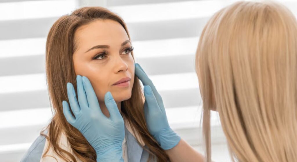
Skin diagnosis often begins with a careful visual examination. When you visit a dermatologist, they will usually look closely at the colour, shape, distribution, and texture of any skin changes. These visible features can provide valuable clues about what may be affecting your skin. A thorough examination helps your dermatologist form an initial understanding of the condition.
However, many skin conditions can appear quite similar at first glance. Issues such as eczema, psoriasis, fungal infections, and certain drug reactions may look alike in their early stages. Because of this, relying only on what can be seen on the surface may not always provide a clear answer. Your dermatologist may therefore need additional information to confirm the diagnosis.
In some situations, your doctor may recommend examining a small sample of your skin under a microscope. This process is known as histopathology. The sample is carefully analysed in a laboratory to observe the structure of the skin cells and tissues. These microscopic details can reveal patterns that are not visible during a standard examination.
When these laboratory findings are considered alongside what your dermatologist observed on your skin, it creates a more complete picture. This combined approach is known as clinicopathological correlation. By linking clinical observations with microscopic analysis, your dermatologist can reach a more accurate diagnosis. For you, this means a better understanding of your condition and a more suitable treatment plan.
What Is Clinicopathological Correlation?
Clinicopathological correlation is the process of linking what your doctor observes on your skin with what appears under the microscope. During your examination, the clinical perspective focuses on the visible signs of your condition, such as colour, texture, and distribution. The pathological perspective, on the other hand, looks at the structure of your skin cells in a tissue sample. When these two viewpoints are considered together, they provide a deeper understanding of what may be affecting your skin.
If only one perspective is used, the diagnosis may not always be clear. The appearance of your skin may suggest several possible conditions. At the same time, microscopic findings from a biopsy can sometimes be difficult to interpret without knowing your symptoms and medical history. Because of this, both types of information are important.
When your dermatologist and pathologist work together to interpret these findings, the diagnosis becomes more reliable. Your clinical history and visible symptoms help guide the laboratory analysis. In turn, the microscopic findings can confirm or refine the doctor’s initial impression. This collaborative approach helps ensure that you receive a more accurate diagnosis and appropriate treatment.
Why Skin Conditions Can Be Difficult to Identify
Skin conditions can sometimes be difficult to identify because many of them share similar features. You may notice symptoms such as redness, scaling, itching, or inflammation, but these signs appear in a wide range of skin diseases. Because of this overlap, it is not always easy for your dermatologist to determine the exact cause based only on what can be seen during an examination. In some cases, additional evaluation is needed to reach a clearer diagnosis.
The appearance of a skin condition can also change over time. Factors such as scratching, infection, or previous treatments may alter how your skin looks. These changes can make the condition harder to recognise. By examining a small skin sample under a microscope, doctors can observe details that are not visible to the naked eye. These microscopic patterns help provide more accurate information about what may be affecting your skin.
The Role of Clinical Examination
When you visit a dermatologist, the clinical examination is usually the first and most important step in identifying a skin condition. Before any laboratory tests are considered, the doctor carefully examines your skin to understand what may be happening.
1. Observing the Pattern and Distribution of Lesions: During a clinical examination, your dermatologist looks closely at how skin lesions appear and where they are located on your body. The pattern, shape, and distribution of these lesions can often provide valuable diagnostic clues. For example, certain conditions tend to appear on specific areas of the body, while others follow recognisable patterns that help narrow down the possible causes.
2. Understanding Your Medical History: Your medical history plays a crucial role in helping the dermatologist interpret what they see on your skin. You may be asked about when your symptoms first appeared, whether they have changed over time, and if you have experienced issues such as itching, pain, or sudden spreading of lesions. This information helps the doctor connect your current symptoms with possible underlying conditions.
3. Considering Lifestyle and Medication Factors: Lifestyle factors and medications can sometimes influence skin health. Your dermatologist may ask about your daily habits, skincare routine, recent illnesses, or any medicines you are taking. In some cases, certain medications or environmental exposures may trigger or worsen skin conditions, making this information essential for accurate diagnosis.
4. Deciding Whether Further Tests Are Needed: The clinical examination helps your dermatologist determine whether additional tests, such as a skin biopsy, are necessary. Not every skin condition requires laboratory investigation, and many can be diagnosed through careful observation alone. However, if the diagnosis is uncertain or a more detailed analysis is needed, further tests may be recommended.
Overall, the clinical examination provides the foundation for accurate dermatological diagnosis. It allows your dermatologist to understand the full context of your skin condition before relying on laboratory findings. When additional investigations are performed, they are interpreted alongside the clinical observations made during your consultation.
Understanding Skin Biopsy
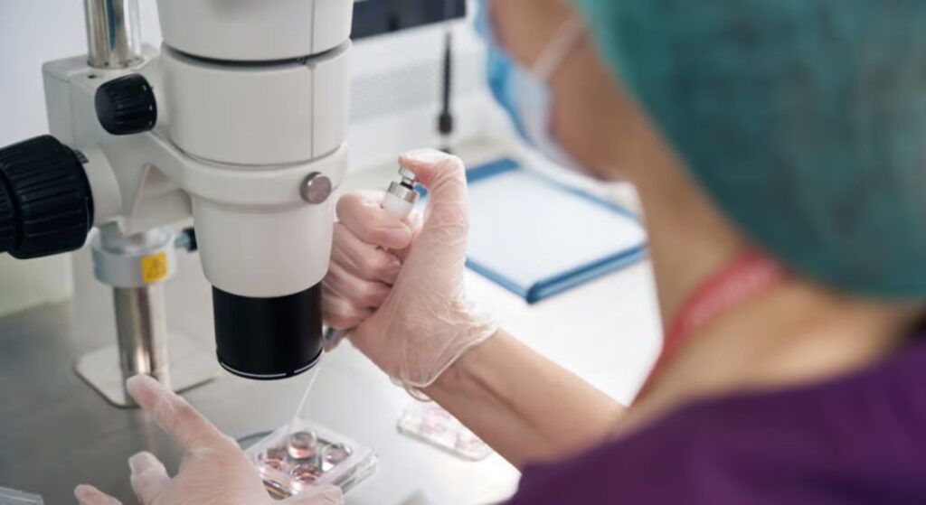
A skin biopsy is a procedure where a small sample of your skin tissue is removed for closer examination. Your dermatologist may recommend this if the diagnosis is not completely clear from a visual assessment alone. The sample is then sent to a laboratory where a specialist, known as a pathologist, studies it under a microscope. This process can provide valuable information about what may be affecting your skin.
The procedure itself is usually quick and straightforward. Your dermatologist will normally use a local anaesthetic to numb the area, so you should feel minimal discomfort. Once the small sample is taken, it is carefully preserved and sent to the laboratory. There, the tissue is processed and very thin sections are placed on slides for detailed examination.
By analysing the sample under a microscope, doctors can see structures that are not visible during a standard skin examination. This includes patterns in the skin cells, the arrangement of tissue layers, and signs of inflammation. These microscopic details help your dermatologist better understand your condition. As a result, you are more likely to receive an accurate diagnosis and the most suitable treatment.
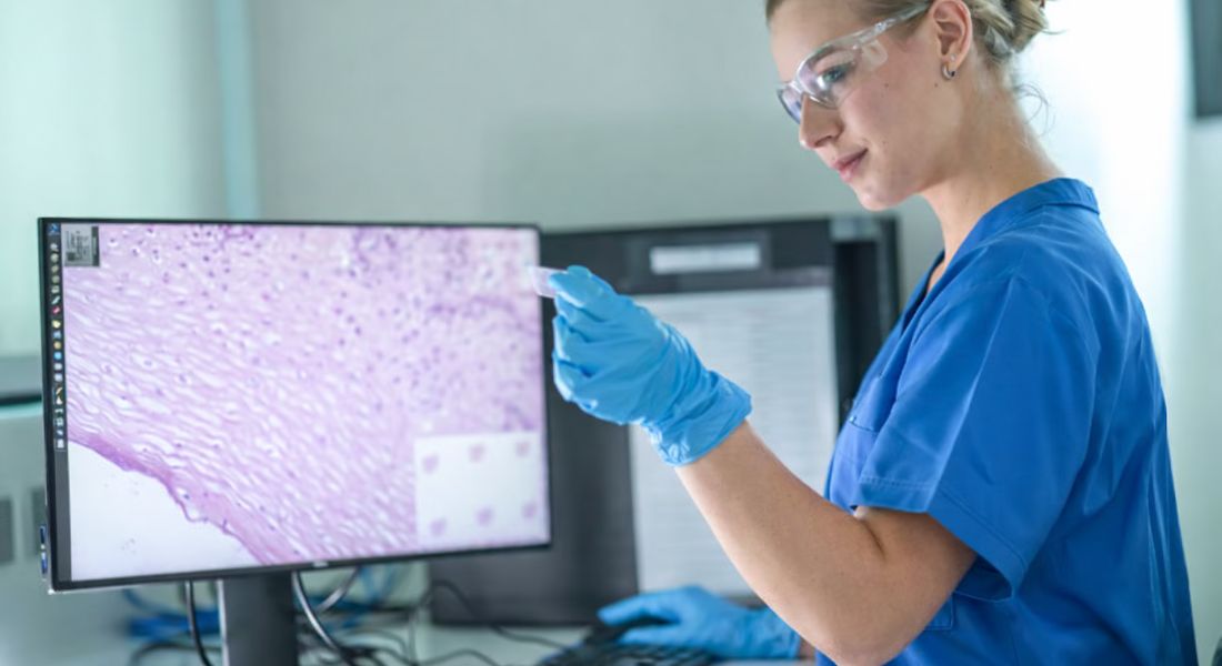
Histopathology in Dermatology
Histopathology refers to the study of tissue structure under a microscope. In dermatology, this analysis focuses on the different layers of your skin and the surrounding cells. By examining these structures closely, specialists can identify subtle changes that may indicate a particular skin condition. This process helps provide a deeper understanding of what may be affecting your skin.
When your skin sample is analysed, the pathologist looks for specific patterns within the tissue. These may include signs of inflammation, unusual cell growth, or areas where the skin structure has been damaged. Certain skin diseases create characteristic microscopic features that can guide the diagnosis. These details help narrow down the possible conditions affecting your skin.
For you, this microscopic analysis can provide valuable information that cannot be seen during a routine examination. It helps your dermatologist understand how the condition is affecting your skin at a cellular level. This insight often supports more accurate treatment decisions. As a result, your care can be better tailored to your specific diagnosis.
However, histopathology alone does not always provide a complete answer. Some microscopic findings can appear similar in different skin conditions. Because of this, the laboratory results must be interpreted alongside your symptoms and clinical examination. When both perspectives are considered together, the diagnosis becomes much clearer.
Communication Between Dermatologist and Pathologist
Effective clinicopathological correlation relies on clear communication between your dermatologist and the pathologist. When a skin biopsy is taken, your dermatologist usually provides important clinical details along with the sample. This information helps the pathologist understand what your skin looked like during the examination. It also guides how the tissue sample is interpreted in the laboratory.
Details such as where the skin lesion is located, how long it has been present, and any symptoms you may be experiencing are often included. In some cases, photographs of your skin condition may also be shared with the laboratory. These images give the pathologist a better idea of the visible changes on your skin. Having this context can make the microscopic findings more meaningful.
After studying the tissue sample, the pathologist sends their findings back to your dermatologist. Your dermatologist then reviews these results alongside your symptoms and clinical examination. By combining both perspectives, your doctor can reach a more accurate diagnosis. This collaborative process helps ensure that you receive the most appropriate treatment for your skin condition.
When Clinical Appearance Is Misleading
Sometimes a skin condition can change its appearance as time passes. Early signs may look quite different from more developed stages, and factors such as treatment, irritation, or infection can also alter how your skin appears. Because of this, relying only on what can be seen during an examination may occasionally be misleading. In certain cases, your dermatologist may recommend a biopsy to look more closely at the skin tissue.
When the sample is examined under a microscope, specific patterns within the cells and tissue can become visible. These details may help clarify what is actually causing the problem. By combining what your dermatologist observes on your skin with the microscopic findings, clinicopathological correlation provides a more reliable diagnosis. For you, this approach helps reduce the risk of misinterpretation and supports more accurate treatment decisions.
Conditions That Often Require Correlation
Some skin conditions often require clinicopathological correlation to reach a clear diagnosis. Autoimmune skin diseases are a good example, as they can create complex patterns that are difficult to confirm through visual examination alone. Conditions such as lupus or lichen planus may require microscopic analysis to understand what is happening within your skin. Looking at both the clinical signs and the tissue sample helps your dermatologist make a more confident diagnosis.
Skin cancers are another situation where this approach is especially important. If a suspicious lesion is found on your skin, your dermatologist may recommend a biopsy. The laboratory analysis can reveal the exact type of tumour and how deeply it extends into the skin. This information is essential for planning the most appropriate treatment for you.
Chronic inflammatory conditions may also benefit from this combined approach. Conditions such as psoriasis, eczema, or certain drug reactions can sometimes appear similar on the surface. By examining a tissue sample alongside your symptoms and medical history, your dermatologist can better understand the condition. This helps ensure that you receive the most suitable treatment for your skin.
Importance in Skin Cancer Diagnosis
Diagnosing skin cancer often relies on careful microscopic evaluation. While your dermatologist may identify a suspicious lesion during a clinical examination, a biopsy is usually needed to confirm the diagnosis. The tissue sample is examined under a microscope to understand the nature of the growth. This step helps ensure that the diagnosis is accurate.
Different types of skin cancer behave in different ways and require different treatments. For example, basal cell carcinoma, squamous cell carcinoma, and melanoma each develop and spread differently. By analysing the tissue sample, specialists can determine the exact type of cancer affecting your skin. This information is essential for deciding the most suitable treatment for you.
Clinicopathological correlation plays a key role in this process. Your dermatologist first identifies any concerning changes during the examination. The microscopic findings then confirm the diagnosis and provide further detail about the tumour. By combining both perspectives, doctors can guide your treatment more effectively and safely.
Identifying Inflammatory Skin Disorders
Inflammatory skin conditions can often look quite similar when you first notice them. Symptoms such as redness, scaling, and itching appear in many different disorders. Because these signs overlap, it can sometimes be difficult for your dermatologist to identify the exact condition based only on what is visible on your skin. In such cases, further investigation may be needed to reach a clearer diagnosis.
By examining a small skin sample under a microscope, doctors can observe specific inflammatory patterns within your skin. Certain immune cells or structural changes may point towards a particular condition. When these microscopic findings are considered alongside your symptoms and medical history, they become much more meaningful. This combined approach helps your dermatologist interpret the results accurately and choose the most suitable treatment for you.
Role in Rare Skin Conditions
When you develop a rare skin condition, the diagnosis may not always be straightforward. Some uncommon diseases present with unusual symptoms or patterns that are difficult to recognise during a routine examination. In these situations, dermatologists often rely on additional investigations to understand what is happening beneath the surface of your skin.
1. Unusual or Atypical Skin Presentations: Rare skin diseases often appear in ways that do not follow common dermatological patterns. When your dermatologist encounters lesions that look unfamiliar or behave differently from typical conditions, it may raise the possibility of a rare disorder.
2. Microscopic Clues from Histopathology: Once a biopsy sample is taken, it is examined under a microscope in a process known as histopathological analysis. This examination can reveal structural changes in the skin cells, layers, and surrounding tissues that are not visible to the naked eye.
3. Use of Special Stains and Additional Tests: In some cases, the tissue sample from your biopsy may undergo further testing using specialised laboratory techniques. These can include special stains or molecular tests designed to highlight particular cells, proteins, or microorganisms. Such methods can help your dermatologist and pathologist detect infections.
4. Combining Clinical and Microscopic Findings: Dermatologists rely on a process known as clinicopathological correlation, which means combining the findings from your clinical examination with the results of the microscopic analysis. By bringing these two perspectives together.
Overall, biopsies and histopathological analysis provide valuable support when diagnosing rare skin conditions. They help dermatologists confirm or rule out unusual diseases that may not be obvious during an initial examination.
Improving Diagnostic Accuracy
When your dermatologist examines your skin, they observe visible patterns, symptoms, and the overall appearance of the condition. At the same time, microscopic analysis of a biopsy can reveal changes within the skin cells and tissue layers. When these two perspectives are considered together, they create a clearer and more complete understanding of what may be affecting your skin.
Clinical observation helps identify possible conditions based on how your skin looks and how your symptoms have developed. Histopathology then confirms whether specific structural or cellular changes are present in the tissue. When the clinical findings and microscopic results support each other, your dermatologist can be much more confident about the diagnosis. This combined approach helps reduce uncertainty.
For you, a more accurate diagnosis means that treatment can be planned more effectively. Instead of relying on trial treatments, your dermatologist can choose therapies that directly target the confirmed condition. This may lead to better results and a more efficient treatment process. Ultimately, it helps improve your overall skin health and treatment outcomes.
Challenges in Correlation
Although clinicopathological correlation is very valuable, it can sometimes present challenges. Some skin conditions produce microscopic features that look quite similar to each other. This can make it difficult for specialists to interpret the tissue findings with complete certainty. In addition, if the clinical information provided with the biopsy is limited, the pathologist may not have enough context to fully understand the results.
This is why clear communication between your dermatologist and the pathologist is so important. When detailed information about your symptoms, medical history, and skin appearance is shared, the laboratory findings become easier to interpret. In some cases, specialists may discuss the results together to review any uncertainties. This collaborative approach helps strengthen diagnostic decisions and ensures you receive the most accurate assessment possible.
Advances in Dermatopathology
Modern technology has significantly improved the field of dermatopathology. Today, digital imaging and advanced staining techniques allow specialists to see tissue structures with much greater clarity. These tools help highlight subtle changes within your skin cells that may not have been easy to detect in the past. As a result, dermatologists and pathologists can work with more detailed information when evaluating your condition.
Molecular testing is also becoming more common in dermatology. By analysing genetic markers within a tissue sample, doctors may be able to identify certain skin diseases more precisely. These tests can provide additional clues about how a condition develops and how it may respond to treatment. For you, this means that diagnosis and treatment planning may become even more targeted and personalised.
Even with these technological advances, clinicopathological correlation remains essential. Technology can support the diagnostic process, but it cannot replace clinical experience and judgement. Your dermatologist still needs to interpret laboratory findings alongside your symptoms and skin examination. When these perspectives are combined, they provide the most reliable path to an accurate diagnosis.
The Importance of Experience
xperience plays a very important role when your skin biopsy is being interpreted. Skilled dermatologists are often able to recognise subtle patterns in the way a skin condition appears. At the same time, experienced pathologists can identify microscopic features within the tissue that might otherwise be overlooked. This combination of expertise helps ensure that small but important details are not missed.
Years of training allow specialists to analyse complex findings with greater confidence. Dermatologists understand how different skin conditions present clinically, while pathologists focus on the cellular changes seen under the microscope. When both perspectives are carefully considered, the diagnosis becomes much more reliable. This shared knowledge strengthens the overall assessment of your condition.
Collaboration between specialists is therefore a key part of dermatological care. Your dermatologist and the pathologist may review findings together to ensure that the clinical and microscopic information align. These discussions help clarify uncertain cases and refine the diagnosis. Working as a team improves the quality of the final decision.
For you as a patient, this expertise can make a significant difference. A more accurate diagnosis allows your dermatologist to choose treatments that are better suited to your condition. Instead of guesswork, your care is guided by specialist knowledge and careful analysis. This approach supports more effective treatment and better long-term outcomes.
Patient Perspective on Skin Biopsy
It is quite common for you to feel anxious if your dermatologist recommends a skin biopsy. However, the procedure is usually quick and straightforward. Your doctor will normally use a local anaesthetic to numb the area, so you should feel very little discomfort during the process. Knowing what to expect can often help you feel more at ease.
Understanding why the biopsy is needed may also reduce your concerns. The small tissue sample helps confirm a diagnosis and guides the most suitable treatment for your skin condition. Most biopsies heal well and leave only minimal scarring when proper aftercare is followed. Your dermatologist will provide clear instructions to support healing, and the valuable information gained from the biopsy often outweighs the temporary inconvenience.
Why Correlation Improves Treatment Outcomes

Accurate diagnosis plays a major role in how successful your treatment will be. When your dermatologist clearly identifies the exact condition affecting your skin, the therapy can be chosen more precisely. This helps avoid treatments that may not be suitable for your condition. As a result, your care can be more focused and effective from the beginning.
If a skin condition is misdiagnosed, it may lead to treatments that do not work as expected. This can sometimes prolong your symptoms and delay improvement. Clinicopathological correlation helps reduce this risk by combining what your dermatologist observes with what is seen under the microscope. Looking at both perspectives together makes the diagnosis more reliable.
For you, a more accurate diagnosis often leads to better treatment outcomes. Once your dermatologist fully understands the nature of your condition, they can select therapies that are better suited to your needs. This approach supports faster improvement and more effective long-term management of your skin health. Ultimately, the goal of clinicopathological correlation is to ensure you receive the most appropriate care possible.
FAQs:
1. What is clinicopathological correlation in dermatology?
Clinicopathological correlation means combining what your dermatologist sees on your skin with what is found when a biopsy sample is examined under a microscope. The clinical examination focuses on visible signs such as colour, shape, and distribution of lesions, while the laboratory analysis studies the skin tissue at a cellular level. When these two perspectives are considered together, the diagnosis becomes clearer and more reliable for you.
2. Why can skin conditions be difficult to diagnose by appearance alone?
Many skin diseases share similar symptoms such as redness, scaling, itching, or inflammation. Because of this, different conditions can look almost the same during a visual examination. In some cases, scratching or previous treatments may also change how your skin appears. For you, this is why dermatologists sometimes recommend additional tests to confirm the diagnosis.
3. What is a skin biopsy and why might it be recommended?
A skin biopsy is a simple procedure where your dermatologist removes a small sample of skin for laboratory analysis. The area is usually numbed with a local anaesthetic, so you should feel minimal discomfort. The tissue is then examined under a microscope to identify patterns that cannot be seen on the surface. For you, this helps confirm the exact cause of the skin problem.
4. How does histopathology help identify skin conditions?
Histopathology involves studying the structure of skin tissue under a microscope. Specialists look for patterns such as inflammation, unusual cell growth, or changes in skin layers. These microscopic clues help doctors understand what may be affecting your skin. For you, this deeper analysis often supports a more accurate diagnosis.
5. Why is communication between the dermatologist and pathologist important?
When a biopsy is sent to the laboratory, your dermatologist provides information about your symptoms and how the skin lesion appears. This context helps the pathologist interpret the tissue sample correctly. Both specialists then review the findings together. For you, this collaboration improves the accuracy of the final diagnosis.
6. Which skin conditions often require clinicopathological correlation?
Some conditions require both clinical examination and microscopic analysis to confirm the diagnosis. Examples include autoimmune diseases, inflammatory skin disorders, and certain drug reactions. Skin cancers also require biopsy confirmation. For you, this combined approach helps ensure the condition is correctly identified.
7. How does this approach help diagnose skin cancer?
When a suspicious lesion is found, a biopsy allows specialists to examine the tissue under a microscope. This analysis helps identify the type of skin cancer and its characteristics. The clinical examination and microscopic findings are then considered together. For you, this ensures the diagnosis is accurate and guides treatment planning.
8. Can skin conditions change in appearance over time?
Yes, skin conditions may change as they develop or respond to treatment. Infection, irritation, or scratching can also alter how your skin looks. Because of these changes, the condition may not always appear typical during examination. For you, microscopic analysis can help clarify the diagnosis.
9. Are skin biopsies generally safe?
Skin biopsies are routine procedures performed in dermatology clinics. A local anaesthetic is used to numb the area before the small sample is taken. Most people experience little discomfort and the area usually heals quickly. For you, the biopsy provides valuable information that supports accurate diagnosis.
10. How does clinicopathological correlation improve treatment decisions?
When clinical observations and microscopic findings support each other, your dermatologist can identify the condition more confidently. This allows treatments to be chosen more precisely for your skin problem. As a result, you are more likely to receive effective care from the start.
Final Thoughts: Seeing Skin Conditions from Every Angle
Clinicopathological correlation highlights how important it is to look at skin conditions from more than one perspective. While a careful clinical examination provides valuable clues about what may be affecting your skin, microscopic analysis can reveal details that are not visible on the surface. When these two viewpoints are combined, your diagnosis becomes far more precise. For you, this approach helps reduce uncertainty and ensures that treatment decisions are based on a deeper understanding of your condition.
This process also reflects the collaborative nature of modern dermatology. Dermatologists and pathologists work together to connect clinical observations with laboratory findings, strengthening the accuracy of the final diagnosis. By linking visible symptoms with cellular changes, specialists can identify skin diseases more reliably and choose treatments that are better suited to your needs.
If you’re thinking about seeing an experienced dermatologist in London, you can contact us at London Dermatology Centre to book a consultation with one of our specialists. Speaking with a specialist ensures your skin concerns are carefully assessed, your diagnosis is supported by detailed clinical evaluation, and your treatment plan is guided by expert dermatological care.
References:
- Malik, A., Siraj, F., Shruti, S., Gupta, P., Khullar, G. and Ramesh, V. (2020) Clinicopathological Concordance in 2216 Cases of Skin Biopsy over One Year: An Indian Experience, Cureus, 12(4), e7752. Available at: https://www.ncbi.nlm.nih.gov/pmc/articles/PMC7243079/
- Talwar, A., Pai Ballambat, S., Rao, R. and Shetty, V.M. (2022) Clinico‑Pathological Correlation in Dermatological Disorders: A Retrospective Audit of 332 Skin Biopsies from a Tertiary Care Center, Indian Dermatology Online Journal, 14(1), pp. 61–66. Available at: https://pubmed.ncbi.nlm.nih.gov/36776159/
- Lee, J. (2012) Clinicopathological consistency in skin disorders: a retrospective study of 3949 pathological reports, Journal of the American Academy of Dermatology, 66(3), pp. 393–400. Available at: https://www.sciencedirect.com/science/article/abs/pii/S019096221100003X
- Tomasini, C. F., Michelerio, A., Isoletta, E., Barruscotti, S., Wade, B. & Muzzi, A. (2023) A Clinico‑Pathological Multidisciplinary Team Increases the Efficacy of Skin Biopsy and Reduces Clinical Risk in Dermatology, Dermatopathology, 10(2), pp. 153–167. Available at: https://www.mdpi.com/2296‑3529/10/2/23
- Wickramasinghe Mudiyanselage S. N., Jayasooriya, P. R., Nihal Mendis, B. R. & Lombardi, T. (2025) Comparative Clinicopathological Analysis of Oral Focal Mucinosis and Solitary Cutaneous Focal Mucinosis: A Case Series and Literature‑Based Analysis, Dermatopathology, 12(4), 38. Available at: https://www.mdpi.com/2296-3529/12/4